Why Accurate Retinal Imaging Matters for Early Glaucoma Detection
Glaucoma can gradually damage the optic nerve without causing noticeable symptoms during its early stages. Some people continue seeing normally even as changes develop inside their eyes, making regular examinations important for protecting long-term vision.
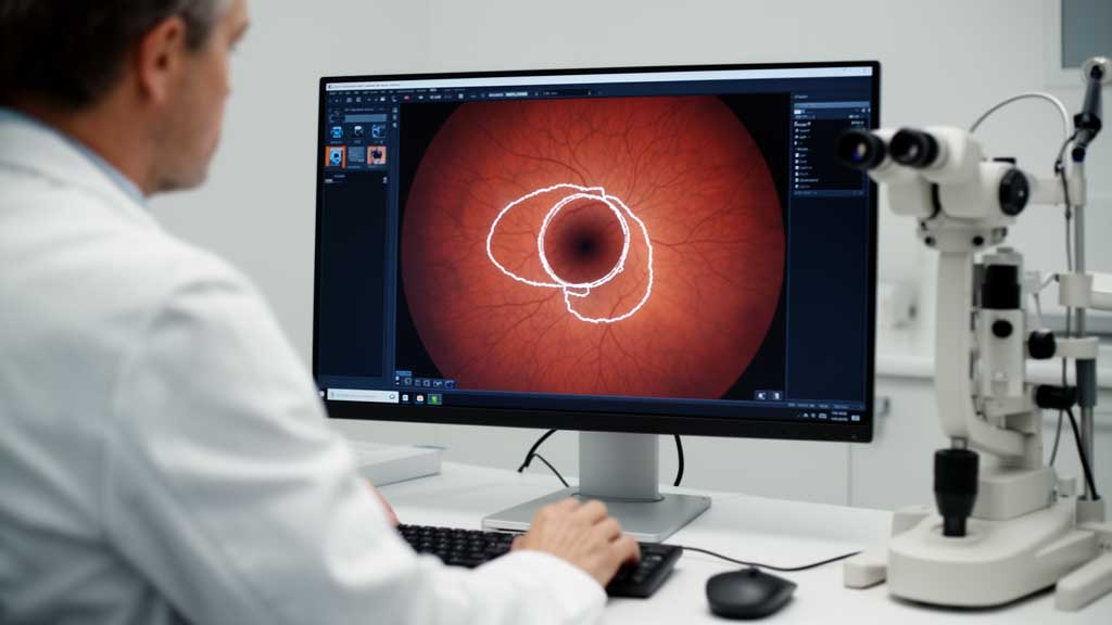
One of the structures eye specialists examine is the optic disc, where nerve fibers leave the back of the eye. Changes in the relationship between the optic disc and optic cup can provide important information when evaluating possible glaucoma.
Researchers are now exploring artificial intelligence (AI) to improve how these structures are identified in retinal photographs. A study published in BMC Ophthalmology in 2026 introduces a deep learning system called EEGA-VMUNet, designed to improve the segmentation of optic disc and optic cup boundaries.
The research combines Vision Mamba architecture with an enhanced edge-guided attention mechanism. Its results demonstrate promising performance across four publicly available retinal image datasets, although additional clinical validation is necessary before the technology can be used independently for diagnosis.
Understanding the Optic Disc and Optic Cup
The optic disc is a circular region at the back of the eye where retinal nerve fibers converge to form the optic nerve. Ophthalmologists examine this area because changes in its appearance can provide clues about possible nerve damage.
Inside the optic disc is a smaller depression called the optic cup. As glaucoma damages retinal ganglion cells and their nerve fibers, the optic cup may become larger relative to the surrounding disc.
Eye specialists commonly evaluate this relationship using the cup-to-disc ratio (CDR). This measurement compares the dimensions of the optic cup with those of the entire optic disc.
An increased cup-to-disc ratio may indicate glaucoma-related changes, but it does not automatically establish a diagnosis. Some healthy individuals naturally have larger optic cups, so specialists consider other findings, including intraocular pressure, retinal nerve fiber thickness, and visual field results.
Accurately identifying the boundaries of these structures is essential when calculating the ratio. Unfortunately, the optic cup can have faint edges, limited contrast, and overlapping retinal blood vessels, making automated measurements challenging.
What Is the EEGA-VMUNet AI Technology?
The research team, led by Lili Cai, Keke Hou, and Zhong Mo, developed EEGA-VMUNet to address difficulties in separating the optic disc and optic cup from surrounding retinal tissue.
Their research is titled “Optic disc and cup segmentation using Vision Mamba UNet with enhanced edge-guided attention in fundus images.” It appeared in BMC Ophthalmology, Volume 26, Issue 1, Article 691, in 2026.
The system builds on Vision Mamba UNet, also known as VM-UNet. This architecture uses a state space model called Mamba to process image information and recognize relationships between different regions.
Unlike conventional transformer approaches that use self-attention, Mamba-based systems are designed to model long-range relationships with computational requirements that scale linearly with input length.
For retinal imaging, this capability allows the network to examine broader anatomical patterns. When the boundary of the optic cup is difficult to distinguish, information about the surrounding disc and the relative positions of both structures can help improve segmentation.
How enhanced edge-guided attention works
The researchers introduced an enhanced edge-guided attention module as the main improvement to the original architecture. It combines global attention with a dual attention mechanism through two parallel processing branches.
The module receives information from the encoder, decoder, and edge feature maps generated through Laplacian pyramid decomposition. These components help the network preserve important spatial details and distinguish anatomical boundaries.
The approach is particularly relevant when retinal blood vessels interfere with the appearance of the optic cup. By incorporating additional edge information, the system aims to reduce segmentation errors around these difficult regions.
Testing also demonstrated the contribution of both attention components. Removing global attention reduced the optic cup Dice score from 92.23% to 91.28%, while removing dual attention lowered it to 91.63% on the evaluated dataset.
How the Researchers Prepared Retinal Images
Fundus photographs contain substantial background information, meaning that analyzing an entire high-resolution image can require unnecessary computing resources.
To address this issue, the team incorporated a YOLOv10 object detection system to locate the optic disc before performing segmentation.
The detector was trained using 400 full-field images from the REFUGE dataset. Once the optic disc was located, the system extracted a smaller region containing the relevant anatomical structures and resized it to 256 by 256 pixels.
A 30% padding margin was applied around the detected region to help ensure that the optic disc remained completely visible in the extracted image.
On the REFUGE validation set, the detector achieved a mean average precision of 0.9944 at an intersection-over-union threshold of 0.5. Precision reached 0.9924, while recall was 0.9788.
The padded cropping procedure successfully included the optic disc in 100% of the REFUGE images evaluated.
Study Results Across Four Retinal Image Datasets
The researchers evaluated EEGA-VMUNet using four publicly available datasets containing retinal photographs with expert-annotated optic disc and optic cup boundaries.
These included Drishti-GS with 101 images, RIM-ONE-r3 with 159 images, ORIGA with 650 images, and REFUGE with 1,200 images.
During five-fold cross-validation on the Drishti-GS training set, the original VM-UNet achieved Dice scores of 96.94% for optic disc segmentation and 90.42% for optic cup segmentation.
Adding the enhanced edge-guided attention module increased these measurements to 97.50% and 92.23%, respectively. Incorporating the multi-scale loss function produced the complete EEGA-VMUNet model, which achieved the strongest overall results across the evaluated metrics.
Comparison with other artificial intelligence models
The researchers compared their approach against 10 competing models, including U-Net, attention-based U-Net variants, transformer-based architectures, MEGANet, Swin-UMamba, and MedSAM.
In Drishti-GS cross-validation, EEGA-VMUNet achieved an optic disc Dice score of 97.73% and an intersection-over-union score of 95.56%. These were the highest reported results among the evaluated models for those measurements.
Performance remained competitive on other datasets. The model achieved an optic disc Dice score of 95.20% on ORIGA and 97.26% on RIM-ONE-r3.
The researchers also conducted zero-shot cross-domain testing, training the model on REFUGE and evaluating it directly on Drishti-GS without additional training. It achieved a disc Dice score of 95.18% and a cup Dice score of 85.13%.
These findings suggest that the architecture can retain useful segmentation performance when applied to images from a different dataset. However, results still vary depending on the characteristics of the retinal photographs.
Can AI Use These Measurements to Screen for Glaucoma?
Accurate segmentation is important, but researchers also wanted to determine whether the measurements produced by the system could help distinguish glaucoma cases from those without the disease.
They used AI-generated cup-to-disc ratios to classify glaucoma against official labels from 400 REFUGE test samples.
The model achieved an area under the receiver operating characteristic curve (AUC) of 0.9566. However, its sensitivity and specificity changed considerably depending on the selected classification threshold.
At a threshold of 0.5, sensitivity reached 97.5%, while specificity was 68.6%. At a threshold of 0.6, sensitivity decreased to 72.5%, whereas specificity increased to 95.8%.
Sensitivity describes how effectively a screening method identifies people who have a condition. Specificity measures how effectively it identifies people who do not have that condition.
This difference is important when designing screening programs. A higher-sensitivity setting may help identify more potential glaucoma cases, although the lower specificity can also produce additional false-positive results requiring further examination.
The researchers stressed that the reported findings do not establish EEGA-VMUNet as a standalone diagnostic system. Professional assessment remains necessary to confirm glaucoma and determine the appropriate clinical approach.
Limitations That Could Affect Clinical Use
Although the reported results are encouraging, the researchers identified several situations where the AI system may produce less reliable measurements.
For example, images showing peripapillary atrophy, which involves changes in tissue surrounding the optic disc, sometimes caused the system to overestimate the optic cup. Tilted optic discs and severely degraded photographs also affected prediction accuracy.
The researchers additionally examined how closely AI-generated cup-to-disc ratios agreed with expert measurements. Intraclass correlation coefficients across the four datasets ranged from 0.841 to 0.946.
The absolute mean biases in Bland-Altman analysis remained below 0.01 across all datasets. However, the 95% limits of agreement ranged from 0.1380 to 0.2163, exceeding the previously reported 0.11 benchmark for differences between human observers.
This limitation is important when monitoring individual patients over time. The researchers indicated that the current measurement precision may not yet be adequate for reliably tracking long-term changes in a person’s glaucoma condition.
Additional research is also needed to evaluate the model across more clinical settings, compare it with other emerging Mamba architectures, and reduce its computational requirements for devices with limited processing capacity.
What This Development Means for Future Eye Care
AI-assisted retinal imaging could eventually help eye care professionals evaluate more photographs and identify suspicious optic nerve changes more efficiently.
Automated segmentation may also support screening programs by providing consistent measurements of important retinal structures. However, its usefulness will depend on validation using diverse patient populations and real-world clinical images.
The research team identified several priorities for further development, including multi-center validation, integration of additional imaging information, and model compression.
One particularly relevant opportunity involves combining fundus photography with optical coherence tomography (OCT). OCT provides detailed cross-sectional images of retinal structures, allowing specialists to examine the retinal nerve fiber layer and other tissues affected by glaucoma.
Combining these imaging methods could provide more comprehensive information than relying on optic disc photographs alone. Nevertheless, further studies are necessary to determine whether this approach improves actual patient outcomes.
When Should You See an Eye Doctor?
Developments in AI should not replace regular eye examinations. Glaucoma can progress without noticeable symptoms, making professional screening important even when your eyesight appears normal.
People with a family history of glaucoma, older adults, and individuals with elevated intraocular pressure should discuss appropriate examination schedules with an eye care professional.
During a comprehensive glaucoma evaluation, an ophthalmologist may perform several tests:
- Tonometry: Measures the pressure inside the eye. Elevated intraocular pressure is an important glaucoma risk factor, although glaucoma can also occur with pressures within the statistically normal range.
- Gonioscopy: Examines the drainage angle inside the eye to determine whether it is open or narrow.
- Dilated optic nerve examination: Allows the specialist to inspect the optic disc for changes associated with nerve damage.
- Optical coherence tomography: Evaluates retinal nerve fiber layer thickness and other structures that may be affected by glaucoma.
- Visual field testing: Checks for areas of reduced peripheral vision that may indicate functional damage.
Seek urgent medical attention if you experience sudden severe eye pain, redness, blurred vision, halos around lights, headache, or nausea. These symptoms may indicate acute angle-closure glaucoma, which requires immediate medical assessment.
Taking the Next Step Toward Protecting Your Vision
The development of EEGA-VMUNet demonstrates how advanced AI architectures may improve the analysis of retinal photographs. Its results across four public datasets highlight the potential of combining Vision Mamba with enhanced attention mechanisms to identify optic disc and optic cup boundaries.
However, promising segmentation scores do not automatically translate into reliable clinical diagnoses. Measurement limitations, difficult retinal images, and the need for broader validation remain important challenges.
For now, the most practical way to protect your eyesight is to maintain regular comprehensive eye examinations and follow your ophthalmologist’s recommendations. If you have a family history of glaucoma or have noticed changes in your vision, schedule an eye assessment rather than waiting for symptoms to become more noticeable.
Research reference: Cai, L., Hou, K., & Mo, Z. (2026). Optic disc and cup segmentation using Vision Mamba UNet with enhanced edge-guided attention in fundus images. BMC Ophthalmology, 26(1), Article 691. DOI: 10.1186/s12886-026-05371-x.