Innovative Glaucoma Diagnostics in Philippine Hospitals
Introduction to Glaucoma and its Impact on Vision
The windows to our world are our eyes, allowing us to witness the beauty around us. But what happens when those windows start to cloud over? Glaucoma, a silent thief of vision, can slowly rob us of this precious gift if left undetected and untreated. In the Philippines, where eye health is a growing concern, hospitals are embracing innovative diagnostic technologies to detect and manage glaucoma at an early stage. Join us as we explore these cutting-edge advancements in glaucoma diagnostics that are revolutionizing eye care in Philippine hospitals! So grab your reading glasses (or contact lenses) and let\’s dive into the fascinating world of glaucoma detection!
Traditional Methods of Diagnosing Glaucoma
Traditional Methods of Diagnosing Glaucoma
Diagnosing glaucoma early on is crucial in preventing further damage to one\’s vision. In the past, traditional methods such as tonometry and visual field testing were commonly used by healthcare professionals to detect glaucoma.
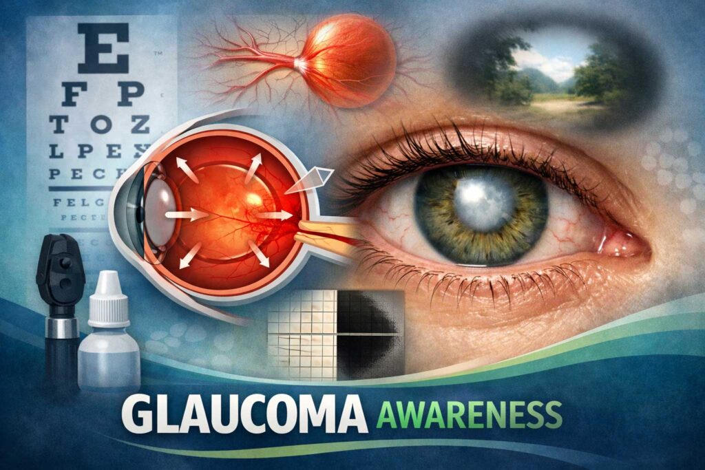
Tonometry involves measuring the pressure inside the eye using a device called a tonometer. Elevated intraocular pressure (IOP) is often associated with glaucoma and can be an important indicator for diagnosis.
Visual field testing, also known as perimetry, assesses the patient\’s peripheral vision. This test measures how well they can see objects in their side or \”peripheral\” vision. Glaucoma typically causes gradual loss of peripheral vision, which can be detected through this examination.
Another method used in diagnosing glaucoma is optic nerve head evaluation. Ophthalmologists examine the optic nerve at the back of the eye for signs of damage or abnormalities indicative of glaucomatous changes.
While these traditional methods have been effective in diagnosing glaucoma to some extent, advancements in technology have revolutionized diagnostic techniques and improved accuracy rates significantly. Let\’s explore these innovative diagnostics next!
Advancements in Glaucoma Diagnostics in Philippine Hospitals
Advancements in Glaucoma Diagnostics in Philippine Hospitals
In recent years, there have been significant advancements in the field of glaucoma diagnostics. These developments have brought about more accurate and efficient methods for detecting this silent thief of sight. Philippine hospitals have also embraced these innovations to provide better care for patients with glaucoma.
One notable advancement is the use of Optical Coherence Tomography (OCT) in glaucoma diagnosis. This non-invasive imaging technique allows doctors to visualize and analyze the optic nerve and retinal nerve fiber layer, which are crucial indicators of glaucoma damage. With OCT, healthcare professionals can detect early signs of the disease and monitor its progression over time.
Another innovative technology being utilized is confocal scanning laser ophthalmoscopy (CSLO). CSLO provides high-resolution images of the optic disc and surrounding structures, aiding in the detection and monitoring of glaucomatous damage.
Additionally, some hospitals now employ visual field testing techniques that utilize sophisticated algorithms to accurately assess a patient\’s peripheral vision. These tests help identify any abnormalities or loss of vision caused by glaucoma.
Moreover, genetic testing has become increasingly important in diagnosing certain types of glaucoma that are associated with specific gene mutations. By identifying these genetic markers, doctors can make more informed decisions regarding treatment options tailored to each patient\’s unique needs.
The advent of telemedicine has also played a role in advancing glaucoma diagnostics in Philippine hospitals. Through remote consultations and virtual follow-ups, patients can receive timely care without having to travel long distances.
These advancements bring several benefits for patients diagnosed with glaucoma – earlier detection means earlier intervention and better preservation of their vision. By utilizing these innovative diagnostic tools and techniques, healthcare providers can offer personalized treatment plans based on an individual\’s specific condition.
While there are challenges involved in implementing these advanced diagnostics across all Philippine hospitals due to cost constraints and limited resources, efforts are being made to make them more accessible. Collaboration between healthcare institutions, government support
The Use of Optical Coherence Tomography (OCT) in Glaucoma Diagnosis
The Use of Optical Coherence Tomography (OCT) in Glaucoma Diagnosis
One innovative technology that has revolutionized the diagnosis and management of glaucoma is Optical Coherence Tomography (OCT). This non-invasive imaging technique allows ophthalmologists to obtain high-resolution cross-sectional images of the optic nerve head and retinal nerve fiber layer.
Using OCT, doctors can accurately measure the thickness of these structures, which are key indicators for diagnosing glaucoma. By comparing these measurements with age-matched normative data, they can identify any signs of thinning or damage caused by elevated intraocular pressure.
What makes OCT particularly valuable is its ability to detect early-stage glaucoma before noticeable vision loss occurs. Traditional methods often rely on visual field tests, which can only detect advanced stages when significant peripheral vision loss has already occurred. With OCT, early detection means earlier intervention and better chances at preserving sight.
Furthermore, OCT enables doctors to track changes over time by performing regular scans. This allows them to monitor disease progression and tailor treatment plans accordingly. Additionally, it helps determine if current treatments are effective or adjustments need to be made.
In conclusion: The use of Optical Coherence Tomography (OCT) in glaucoma diagnosis offers significant advantages in terms of early detection and monitoring progression. Its ability to provide detailed structural information aids ophthalmologists in making accurate diagnoses and implementing timely interventions for improved patient outcomes
Other Innovative Technologies and Techniques for Glaucoma Detection
Other Innovative Technologies and Techniques for Glaucoma Detection:
1. Visual Field Testing: This technique assesses the peripheral vision of a patient by measuring their ability to detect flashing lights at various points in their field of view. It helps in detecting any areas of vision loss, which is an early sign of glaucoma.
2. Electroretinography (ERG): ERG measures the electrical activity generated by the retina when it responds to light stimulation. It can help identify abnormalities in retinal function before any visible damage occurs, allowing for early detection and intervention.
3. Confocal Scanning Laser Ophthalmoscopy (CSLO): CSLO uses laser technology to create high-resolution images of the optic nerve head and surrounding structures. By analyzing these images, doctors can identify subtle changes indicative of glaucoma progression.
4. Corneal Hysteresis Measurement: This non-invasive technique measures the cornea\’s resistance to stress or deformation caused by an applied force. Lower corneal hysteresis has been found to be associated with higher risk for developing glaucoma, making this a valuable diagnostic tool.
5. Genetic Testing: Advances in genetic research have led to the identification of certain gene mutations that increase an individual\’s susceptibility to glaucoma. Genetic testing can help determine if someone carries these mutations and thus may be at higher risk for developing the disease.
These innovative technologies and techniques provide healthcare professionals with additional tools for accurate diagnosis and monitoring of glaucoma patients, ensuring timely intervention when needed
Benefits of Early Detection and Treatment
Early detection and treatment of glaucoma can bring numerous benefits to individuals at risk. By identifying the condition in its early stages, it becomes possible to implement appropriate interventions and prevent further damage to the optic nerve.
One of the key advantages of early diagnosis is preserving vision. Glaucoma is often referred to as a \”silent thief\” because it progresses gradually without causing noticeable symptoms until significant vision loss occurs. Detecting glaucoma before symptoms manifest allows for prompt intervention, which can slow down or halt disease progression, ultimately preventing blindness.
Additionally, early treatment provides an opportunity for better management of intraocular pressure (IOP). Elevated IOP is one of the primary risk factors for glaucoma development and progression. By starting treatment early on, healthcare professionals can work towards achieving target IOP levels more effectively.
Moreover, timely diagnosis enables patients to explore various treatment options available. Depending on individual circumstances and disease severity, treatments may include medications, laser therapy or surgical procedures such as trabeculectomy or drainage implantation. Early detection increases the likelihood of successful outcomes with these interventions.
Furthermore, identifying glaucoma at an earlier stage also helps reduce healthcare costs associated with managing advanced cases. The financial burden related to treating severe vision impairment caused by advanced glaucoma can be substantial compared to regular eye exams that enable preventive measures.
Recognizing and addressing glaucoma in its initial phases through regular screenings offers significant advantages in terms of preserving visual function and quality of life while minimizing potential complications associated with late-stage disease progression.
Challenges and Limitations of Implementing Innovative Diagnostics in Philippine Hospitals
Challenges and Limitations of Implementing Innovative Diagnostics in Philippine Hospitals
Implementing innovative diagnostics for glaucoma in Philippine hospitals comes with its fair share of challenges and limitations. One major hurdle is the cost associated with acquiring and maintaining advanced diagnostic equipment. These technologies can be expensive to purchase, install, and maintain, making it difficult for some hospitals to allocate funds towards these advancements.
Another challenge lies in the training and expertise required to operate these sophisticated devices. Specialized skills are needed to interpret the results accurately and make informed decisions regarding patient care. Ensuring that healthcare professionals receive adequate training can pose logistical challenges within busy hospital settings.
Infrastructure limitations also play a role in implementing innovative diagnostics. Some hospitals may lack the necessary infrastructure or resources needed to support cutting-edge technologies like optical coherence tomography (OCT) machines or other advanced diagnostic tools.
Additionally, there may be regulatory hurdles or bureaucratic processes that slow down the adoption of new diagnostic techniques within healthcare systems. This could result in delays in providing patients with timely access to innovative glaucoma diagnostics.
Despite these challenges, it is essential for Philippine hospitals to strive towards incorporating innovative diagnostics into their practices. By overcoming these obstacles through collaborations with industry partners, government support, and investment in training programs, more patients can benefit from early detection and improved treatment outcomes for glaucoma.
In conclusion…
Conclusion: Impro
Conclusion: Impro
Innovations in glaucoma diagnostics have revolutionized the way this vision-threatening condition is detected and managed in Philippine hospitals. Traditional methods of diagnosing glaucoma, such as tonometry and visual field testing, are still widely used but can sometimes be limited in their accuracy.
Fortunately, advancements in technology have paved the way for more precise and reliable diagnostic tools. One such tool is Optical Coherence Tomography (OCT), which uses light waves to create detailed images of the eye\’s structures. This non-invasive procedure allows healthcare professionals to accurately measure the thickness of the optic nerve fiber layer, a key indicator for glaucoma.
But OCT is just one example among many innovative technologies being utilized in Philippine hospitals. Other techniques like confocal scanning laser ophthalmoscopy (CSLO) and scanning laser polarimetry (SLP) are also proving beneficial for early detection and monitoring of glaucoma progression.
The benefits of early diagnosis cannot be overstated when it comes to managing glaucoma effectively. By catching this chronic disease at its earliest stages, healthcare providers can implement appropriate treatment plans that aim to slow down or halt further vision loss. Regular monitoring with these advanced diagnostic tools ensures that any changes in intraocular pressure or optic nerve health are promptly addressed.
However, there are challenges associated with implementing these innovative diagnostics across all Philippine hospitals. Limited access to specialized equipment and trained personnel may hinder widespread adoption. Additionally, cost considerations may pose barriers to patients seeking these advanced tests.
Despite these limitations, it is clear that innovations in glaucoma diagnostics provide significant advantages over traditional methods. The ability to detect subtle changes within the eye allows for earlier intervention and improved patient outcomes.
In conclusion (without using those exact words), embracing technological advancements such as OCT alongside other innovative techniques will play a crucial role in combating glaucoma-related blindness across the Philippines. By investing in training programs for healthcare professionals and ensuring equitable access to these diagnostic tools, the country can make great strides in preserving