Key Innovations in Glaucoma Detection Techniques
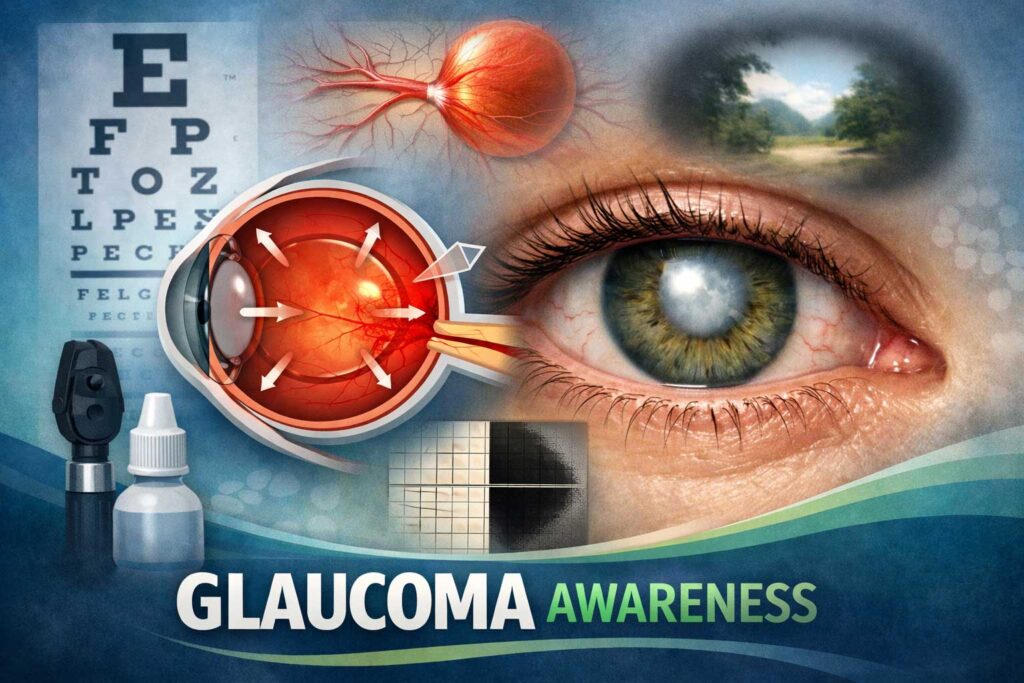
Introduction to Glaucoma
Unlocking the Secrets of Glaucoma: Discover the Future of Eye Care!
Imagine a world where we can detect and treat glaucoma, one of the leading causes of blindness, with unparalleled precision. A world where innovative technologies revolutionize how we diagnose this silent thief that slowly deteriorates our vision. Well, dear readers, that future is already here! In this blog post, we will delve into the fascinating realm of glaucoma detection techniques and explore how cutting-edge advancements are reshaping the landscape of eye care as we know it.
But before we embark on this enlightening journey, let’s take a moment to understand what exactly glaucoma is and why it poses such a grave threat to our precious eyesight. So grab your reading glasses (if you need them) and let’s dive in!
Traditional Diagnostic Techniques for Glaucoma
Traditional Diagnostic Techniques for Glaucoma
Detecting glaucoma at an early stage is crucial to prevent irreversible vision loss. Traditionally, ophthalmologists have relied on a combination of tests to diagnose this condition. One such test is tonometry, which measures the pressure inside the eye. Another common method is visual field testing, which assesses peripheral vision.
Tonometry involves using a device called a tonometer to measure intraocular pressure (IOP). Elevated IOP is often associated with glaucoma and can be an indication of its presence. Visual field testing, on the other hand, evaluates a patient’s ability to see objects in their peripheral vision by presenting stimuli in different areas of their visual field.
Another traditional technique is gonioscopy, which examines the drainage angle of the eye using a special lens. This helps determine if there are any blockages that may contribute to increased intraocular pressure.
Additionally, ophthalmoscopy plays a role in diagnosing glaucoma by allowing doctors to examine the optic nerve for signs of damage or changes indicative of glaucomatous damage.
While these techniques have been valuable tools in diagnosing glaucoma over the years, they do have limitations that can lead to missed diagnoses or delayed treatment initiation. That’s where innovative detection techniques come into play!
Stay tuned as we explore some key innovations revolutionizing how we detect and manage glaucoma!
Limitations of Traditional Techniques
Traditional diagnostic techniques for glaucoma have been widely used for many years, but they do come with some limitations. One of the main drawbacks is that these techniques often rely on subjective assessments by healthcare professionals. This can lead to variations in diagnosis and potentially result in misdiagnosis or delayed treatment.
Another limitation is that traditional techniques primarily focus on measuring intraocular pressure (IOP) as a key indicator of glaucoma. While elevated IOP is a risk factor for developing the condition, it does not always guarantee its presence. As a result, relying solely on IOP measurements may miss cases where other factors contribute to the development of glaucoma.
Furthermore, traditional methods such as visual field testing can be time-consuming and require significant patient cooperation. This can make it difficult to obtain accurate results, especially in patients who may struggle with understanding or following instructions during the test.
Additionally, some traditional techniques are invasive and uncomfortable for patients. For example, gonioscopy involves placing a special lens directly onto the eye’s surface to examine the drainage angle. This procedure can cause discomfort and anxiety among patients.
While traditional diagnostic techniques have played an important role in identifying glaucoma over the years, their limitations highlight the need for new innovations that overcome these challenges and provide more accurate and efficient detection methods.
Key Innovations in Glaucoma Detection
Key Innovations in Glaucoma Detection Techniques
Detecting glaucoma early is crucial for effective treatment and preserving vision. Traditional diagnostic techniques have their limitations, but thanks to key innovations in glaucoma detection, we now have more accurate and efficient methods at our disposal.
One such innovation is Optical Coherence Tomography (OCT), a non-invasive imaging technique that provides detailed cross-sectional images of the retina. By analyzing the thickness of the retinal nerve fiber layer, OCT can detect even subtle changes associated with glaucoma before symptoms manifest.
Another groundbreaking technology is Heidelberg Retinal Tomography (HRT), which uses laser scanning to create three-dimensional images of the optic nerve head. This allows for precise measurement of important parameters like cup-to-disc ratio, enabling early detection and monitoring of glaucomatous damage.
Ultrasound Biomicroscopy (UBM) takes innovation a step further by utilizing high-frequency sound waves to visualize structures behind opaque tissues. With UBM, ophthalmologists can assess anterior chamber angle morphology and identify any abnormalities that may contribute to glaucoma development.
These new techniques offer significant advantages over traditional methods. They provide more accurate measurements, enable earlier detection, improve monitoring capabilities, and enhance treatment planning for patients with glaucoma.
In conclusion (sorry!), these key innovations in glaucoma detection are revolutionizing how we diagnose and manage this sight-threatening disease. As technology continues to advance rapidly, it holds great promise for improving patient outcomes and reducing the burden of vision loss caused by glaucoma.
Optical Coherence Tomography (OCT)
Optical Coherence Tomography (OCT) is a cutting-edge technology that has revolutionized the way glaucoma is detected and monitored. By using light waves to create detailed images of the retina, OCT provides an unprecedented level of accuracy in diagnosing this eye condition.
One of the key advantages of OCT is its ability to capture high-resolution cross-sectional images of the retina. This allows doctors to visualize the thickness of retinal layers and detect any abnormalities or damage caused by glaucoma. Additionally, OCT can measure nerve fiber layer thickness, which is crucial for assessing disease progression.
Another noteworthy feature of OCT is its non-invasive nature. Unlike traditional techniques like gonioscopy or tonometry, which require physical contact with the eye, OCT uses harmless light waves to obtain information about the structure and health of the eye.
Furthermore, OCT offers real-time imaging capabilities, allowing doctors to monitor changes in retinal thickness over time. This enables early detection of glaucoma-related damage even before symptoms become apparent.
In conclusion/Overall/Finally/Optical Coherence Tomography (OCT) represents a significant advancement in glaucoma detection techniques due to its ability to provide precise measurements and detailed images without invasive procedures. With further research and development, this innovative tool has promising potential for improving diagnosis accuracy and enhancing treatment strategies for patients with glaucoma
Heidelberg Retinal Tomography (HRT)
Heidelberg Retinal Tomography (HRT) is a cutting-edge technology that has revolutionized the detection of glaucoma. This non-invasive imaging technique allows for detailed analysis of the optic nerve and retinal structures, providing valuable insights into the progression of this sight-threatening disease.
Using a scanning laser ophthalmoscope, HRT captures high-resolution 3D images of the eye’s posterior segment. By measuring parameters such as optic disc topography, cup-to-disc ratio, and retinal nerve fiber layer thickness, HRT enables clinicians to accurately assess structural changes associated with glaucoma.
One of the key advantages of HRT is its ability to detect early signs of glaucomatous damage even before symptoms become apparent. It facilitates early intervention and treatment planning, significantly improving patient outcomes.
Moreover, HRT offers objective measurements that reduce inter-observer variability commonly encountered in traditional diagnostic techniques. This enhances accuracy and reliability in diagnosing and monitoring glaucoma progression over time.
With continuous advancements in technology, HRT continues to evolve and provide more comprehensive information about glaucomatous changes in the eye. Its integration with other modalities like Optical Coherence Tomography (OCT) further enhances diagnostic capabilities.
Heidelberg Retinal Tomography (HRT) plays a vital role in modern glaucoma detection by providing detailed structural information about the optic nerve head and retina. Its ability to detect subtle changes at an early stage makes it an invaluable tool for timely diagnosis and effective management strategies. The future holds promising possibilities for further innovation in this field as we strive towards better understanding and combating this debilitating condition.
Ultrasound Biomicroscopy (UBM)
Ultrasound Biomicroscopy (UBM) is a cutting-edge technology that has revolutionized the detection and management of glaucoma. This non-invasive imaging technique allows for detailed visualization of the structures within the eye, providing valuable information about the anatomy and function of the anterior segment.
Unlike traditional techniques, UBM utilizes high-frequency ultrasound waves to create high-resolution images of ocular structures. By capturing cross-sectional images, UBM enables clinicians to assess important parameters such as angle width, iris configuration, and ciliary body morphology. This comprehensive evaluation helps in identifying abnormalities associated with glaucoma.
One of the key advantages of UBM is its ability to visualize anatomical features that are difficult to assess using other methods. For instance, it can accurately measure anterior chamber depth and detect abnormalities in trabecular meshwork – crucial factors contributing to increased intraocular pressure in glaucoma patients.
Moreover, UBM provides real-time dynamic imaging capabilities which allow clinicians to observe important physiological changes occurring during eye movements or during different phases of aqueous humor flow. This helps in better understanding disease progression and optimizing treatment strategies tailored specifically for each patient’s needs.
In addition to aiding diagnosis and monitoring progress, UBM also plays a crucial role in preoperative planning for glaucoma surgeries by providing accurate measurements required for surgical interventions like implantation or drainage devices.
Ultrasound Biomicroscopy (UBM) offers an advanced approach to glaucoma detection by providing detailed anatomical information not easily obtainable through traditional methods alone. Its ability to visualize complex structures within the eye makes it an indispensable tool for clinicians striving towards optimal treatment outcomes for their patients with glaucoma.
Comparison of New Techniques with Traditional Ones
Comparison of New Techniques with Traditional Ones
When it comes to the detection and diagnosis of glaucoma, traditional techniques have long been the go-to methods for ophthalmologists. These include tonometry, which measures intraocular pressure, and visual field testing, which assesses peripheral vision. While these methods have proven effective to a certain extent, they do have their limitations.
Enter the new wave of innovative glaucoma detection techniques. One such technique is Optical Coherence Tomography (OCT), which provides high-resolution cross-sectional images of the retina and optic nerve head. This non-invasive imaging technology allows for early detection and monitoring of structural changes associated with glaucoma.
Another groundbreaking technique is Heidelberg Retinal Tomography (HRT). Using laser scanning technology, HRT creates three-dimensional images that help analyze the thickness of retinal nerve fiber layers. This aids in identifying progressive damage caused by glaucoma.
Ultrasound Biomicroscopy (UBM) is another valuable tool in detecting glaucoma-related abnormalities. UBM uses high-frequency sound waves to visualize structures within the eye that are difficult to see with conventional imaging techniques.
Compared to traditional techniques, these new innovations offer several advantages. They provide more precise measurements and detailed imaging of key ocular structures involved in glaucoma progression. They enable earlier detection of subtle changes that may indicate the development or progression of this sight-threatening condition.
Moreover, these advanced technologies allow for better monitoring over time through repeatable measurements and objective data analysis. They also reduce subjectivity in interpretation as many traditional tests rely on subjective patient responses or operator judgment.
The future implications of innovative glaucoma detection techniques are promising indeed! As technology continues to evolve at an exponential rate, we can expect even more accurate diagnostics tools becoming available for ophthalmologists worldwide.
In conclusion,
It’s clear that modern advancements in glaucoma detection have revolutionized how this condition is diagnosed. These new techniques offer greater precision, earlier detection, and improved
Benefits and Future Implications of Innovative Glaucoma Detection
Benefits and Future Implications of Innovative Glaucoma Detection
The development of new techniques for glaucoma detection has brought about numerous benefits and promising future implications. One notable innovation is Optical Coherence Tomography (OCT), a non-invasive imaging test that provides detailed cross-sectional images of the retina. With OCT, doctors can accurately measure the thickness of the optic nerve fiber layer, helping to detect early signs of glaucoma.
Another exciting advancement is Heidelberg Retinal Tomography (HRT), which uses scanning laser technology to create 3D images of the optic nerve head. This allows for precise measurements and analysis, aiding in early diagnosis and monitoring disease progression.
Ultrasound Biomicroscopy (UBM) is yet another groundbreaking technique that utilizes high-frequency sound waves to capture detailed images of the anterior segment structures in the eye. UBM enables clinicians to assess angle structures and identify any abnormalities or changes associated with glaucoma.
These innovative detection techniques offer several advantages over traditional methods. They provide more accurate and detailed information, leading to earlier diagnosis and intervention. Additionally, they are non-invasive procedures that are well-tolerated by patients, minimizing discomfort during testing.
Looking ahead, these innovations hold immense potential for improving patient outcomes in glaucoma care. Early detection means timely treatment initiation, reducing vision loss caused by this progressive disease. Furthermore, these technologies have opened up avenues for ongoing research into better understanding glaucoma etiology and developing targeted therapies tailored to individual patients.
In conclusion,
the advancements we’ve seen in glaucoma detection techniques have revolutionized our ability to diagnose this sight-threatening condition at its earliest stages. With OCT, HRT, UBM, we now have powerful tools at our disposal that enable us to make more informed decisions regarding treatment plans while ensuring greater patient comfort throughout their diagnostic journey.
Conclusion
Conclusion
The field of glaucoma detection has witnessed significant advancements in recent years. Traditional diagnostic techniques, such as tonometry and visual field testing, have played a crucial role in identifying this silent thief of vision. However, these methods have their limitations and may not always provide an accurate assessment of the disease progression.
Fortunately, innovative technologies like Optical Coherence Tomography (OCT), Heidelberg Retinal Tomography (HRT), and Ultrasound Biomicroscopy (UBM) have emerged as valuable tools in the early detection and monitoring of glaucoma. These techniques offer higher precision and detailed imaging of the optic nerve head and retinal layers, allowing for better identification of structural changes associated with glaucoma.
By leveraging these advanced detection methods, ophthalmologists can make more informed decisions regarding treatment plans tailored to individual patients’ needs. Early diagnosis combined with appropriate interventions can significantly slow down or even prevent further vision loss caused by glaucoma.
The benefits offered by these innovations extend beyond improved accuracy in diagnosing glaucoma. They also facilitate proactive management strategies that help maintain optimal ocular health for individuals at risk or those already diagnosed with the condition. Moreover, ongoing research continues to explore new avenues for enhancing our understanding of the etiology of glaucoma, which may pave the way for future breakthroughs in treatment options.
It is important to note that while technology has revolutionized glaucoma detection techniques, regular eye exams remain essential for timely diagnosis and effective management. If you experience any symptoms like blurred vision or increased pressure within your eyes or are at risk due to family history or other factors, it is vital to consult an eye care professional promptly.
As we move forward into a future where advancements continue to shape medical practices around us, it is reassuring to know that scientists and researchers are dedicatedly working towards improving our ability to detect and treat diseases like glaucoma. With continued innovation and collaboration, we can hope to better protect our precious gift of sight and ensure