Army Hospital Pioneers 3D Microscope for Glaucoma Surgery Breakthroughs
In a groundbreaking development, an Army hospital has introduced a cutting-edge 3D microscope to revolutionize glaucoma surgery. This innovative technology promises to enhance precision, improve surgical outcomes, and reduce recovery times for patients suffering from this sight-threatening condition. With glaucoma affecting millions worldwide, this advancement could be a game-changer in ophthalmology.
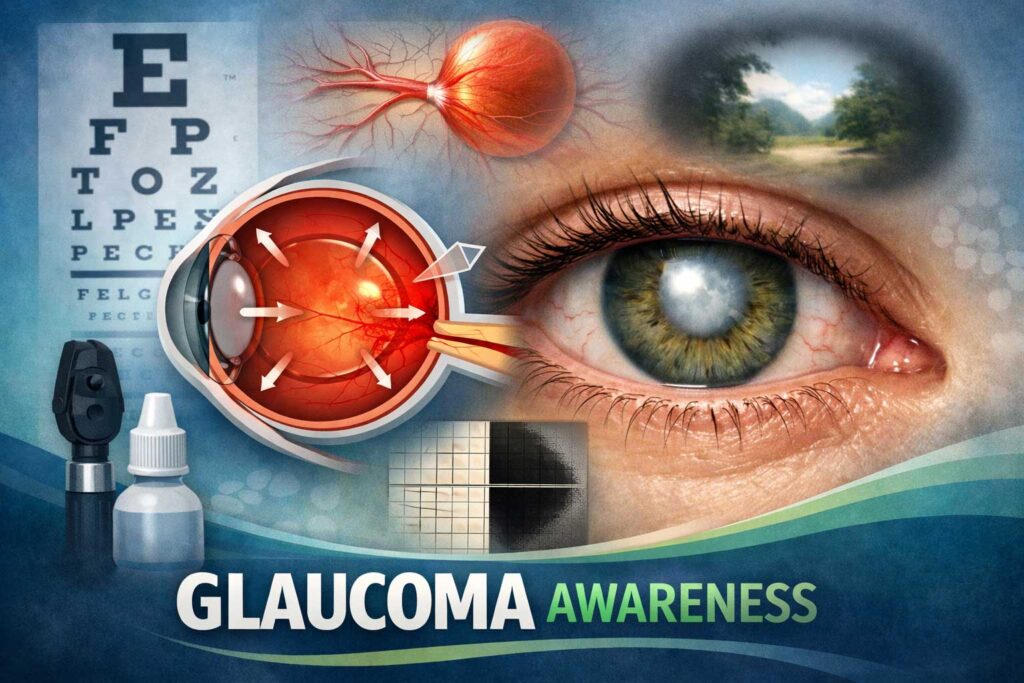
The Rising Challenge of Glaucoma
Glaucoma is one of the leading causes of irreversible blindness, affecting over 80 million people globally. The condition damages the optic nerve, often due to increased intraocular pressure, and requires timely intervention to prevent vision loss. Traditional surgical methods, while effective, come with limitations in precision and visualization.
Surgeons have long relied on conventional microscopes, which provide a two-dimensional view of the surgical field. However, depth perception is critical in delicate eye surgeries, and the lack of 3D visualization has been a persistent challenge.
How the 3D Microscope is Changing the Game
The Army hospital’s adoption of a high-definition 3D microscope marks a significant leap forward. This advanced imaging system offers:
- Enhanced Depth Perception: Surgeons can now see intricate eye structures in three dimensions, improving accuracy during procedures.
- Real-Time Adjustments: The microscope provides dynamic imaging, allowing surgeons to make immediate corrections during surgery.
- Improved Training: The 3D visualization aids in training new surgeons by offering a clearer, more immersive view of surgical techniques.
Key Benefits for Glaucoma Patients
Patients undergoing glaucoma surgery with this technology can expect:
- Faster Recovery: Greater precision means less tissue damage and quicker healing.
- Reduced Complications: Better visualization minimizes risks such as bleeding or unintended damage to surrounding tissues.
- Higher Success Rates: Early studies suggest improved surgical outcomes compared to traditional methods.
The Science Behind the Innovation
The 3D microscope utilizes stereoscopic imaging, which mimics human binocular vision. By capturing two slightly offset images and combining them, the system creates a lifelike three-dimensional view. This is particularly beneficial in glaucoma surgeries, where surgeons work on microscopic structures like the trabecular meshwork.
Additionally, the microscope integrates with augmented reality (AR) overlays, providing real-time data on intraocular pressure and other critical metrics. This fusion of imaging and data analytics ensures that surgeons have all the necessary information at their fingertips.
Case Study: Early Success Stories
Preliminary trials at the Army hospital have shown remarkable results. In one case, a patient with advanced glaucoma underwent a minimally invasive glaucoma surgery (MIGS) using the 3D microscope. The surgeon reported unparalleled clarity, enabling precise placement of a micro-stent to relieve pressure.
Another notable success involved a complex revision surgery where traditional methods had previously failed. The 3D visualization allowed the surgical team to navigate scar tissue and correct the issue without complications.
The Future of Glaucoma Treatment
This breakthrough is just the beginning. Experts predict that as the technology becomes more widespread, it could:
- Lower Healthcare Costs: Fewer complications and repeat surgeries mean reduced expenses for patients and hospitals.
- Expand Access: Portable versions of the 3D microscope could bring advanced care to remote and underserved areas.
- Inspire Further Innovations: The success of this technology may lead to its adaptation in other ophthalmic and even non-ophthalmic surgeries.
Challenges and Considerations
While the benefits are clear, there are hurdles to overcome:
- Cost: High-end 3D microscopes require significant investment, which may limit initial adoption.
- Training: Surgeons must undergo specialized training to maximize the technology’s potential.
- Integration: Hospitals need to upgrade their infrastructure to support these advanced systems.
Conclusion
The Army hospital’s pioneering use of a 3D microscope in glaucoma surgery represents a monumental step forward in eye care. By enhancing precision, reducing risks, and improving patient outcomes, this technology has the potential to redefine how glaucoma is treated worldwide. As research continues and adoption grows, we may soon see this innovation become the gold standard in ophthalmic surgery.
For patients and surgeons alike, the future of glaucoma treatment has never looked brighter—or clearer.