How a Routine Eye Test Saved a Life by Detecting a Brain Tumor
We often think of an eye exam as a simple check for glasses or a screening for common conditions like cataracts. It’s a routine part of healthcare, sometimes even postponed when life gets busy. But what if that 30-minute appointment could do more than just assess your vision? What if it could peer into the very wiring of your brain and uncover a hidden, life-threatening condition? This isn’t a hypothetical scenario. For one patient, a standard visual field test at their optometrist’s office became the critical first step in diagnosing a serious brain tumor, ultimately setting them on a path to treatment and recovery.
This remarkable story underscores a vital truth: your optometrist is often a frontline detective for your overall neurological health. The eyes are not just windows to the soul; they are direct windows to the brain, and subtle changes in vision can be the first and only sign of something much more serious.
The Critical Link: Your Eyes and Your Brain
To understand how an eye test can reveal a brain issue, we need to look at the anatomy. The optic nerves are essentially bundles of over a million nerve fibers that carry visual information from the retina to the brain. They are physically connected to the brain, and any pressure or disruption along this pathway will manifest as changes in vision.
When a tumor grows in or near the pituitary gland, the optic chiasm (where the nerves cross) is a common site of compression. This pressure doesn’t typically cause dramatic blindness overnight. Instead, it creates very specific, gradual deficits in the peripheral vision—a loss that you might not even consciously notice until it’s significant.
The Unsung Hero: The Visual Field Test
This is where the humble visual field test becomes a powerhouse of diagnostic information. During this non-invasive test, you focus on a central point while lights of varying brightness flash in your peripheral vision. You click a button each time you see a flash. The machine maps your complete field of view, creating a detailed chart of your visual sensitivity.
For a patient with a pituitary tumor pressing on the optic chiasm, the resulting pattern is often textbook: a bitemporal hemianopia. This means the outer halves of both the right and left visual fields are missing. It’s a pattern that screams “neurological issue” to a trained eye.
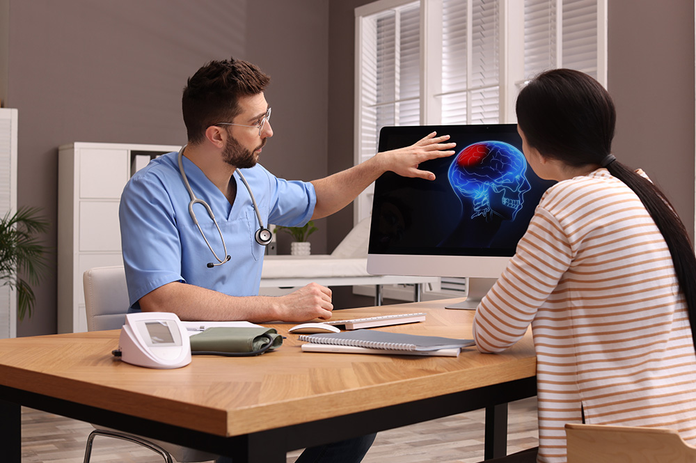
In the case highlighted, the optometrist didn’t just note a slight prescription change. They saw this distinct, alarming pattern on the visual field printout. Recognizing it as a potential red flag for a non-optical problem, they immediately referred the patient to a neurologist and an ophthalmologist for further investigation. That swift action was the pivotal moment.
Beyond the Tumor: What Your Optometrist Can Detect
While brain tumors are a dramatic example, routine eye exams can detect signs of numerous other systemic and neurological conditions. An optometrist examining the retina (the back of the eye) can see blood vessels and nerves directly. This view can reveal:
- Stroke Risk: Evidence of mini-strokes (Hollenhorst plaques) or narrowed retinal arteries can indicate cardiovascular disease and a high risk of a major stroke.
- Multiple Sclerosis (MS): Inflammation of the optic nerve (optic neuritis) is a common early symptom of MS.
- High Blood Pressure: Hypertensive retinopathy shows as damaged, narrowed, or leaking blood vessels in the retina.
- Diabetes: Diabetic retinopathy, characterized by bleeding and fluid leakage in the retina, is a major cause of blindness and is often first detected in an eye exam.
- High Cholesterol: Deposits can appear as yellowish rings around the cornea or as plaques in the retinal blood vessels.
Don’t Ignore the Signs: Symptoms That Warrant a Check-Up
You shouldn’t wait for your scheduled annual exam if you experience sudden or progressive changes in your vision. Be alert to these potential warning signs:
- Unexplained blurring or loss of vision, especially in one eye or a specific part of your visual field.
- Seeing flashing lights, floaters, or shadows that are new or worsening.
- Persistent headaches, particularly if they are new, severe, or accompanied by vision changes.
- Double vision (diplopia).
- Loss of peripheral vision – feeling like you’re looking through a tunnel.
- A drooping eyelid or changes in pupil size.
If you experience any of these, schedule an appointment with your optometrist immediately. Clearly describe your symptoms, including when they started and how they have changed.
A Powerful Reminder for Proactive Health
The story of a life saved by a visual field test is not just a lucky break; it’s a testament to the sophisticated role of modern optometry in holistic healthcare. It highlights why regular, comprehensive eye exams are a non-negotiable part of maintaining your overall well-being, not just your eyesight.
Your optometrist is equipped with the technology and training to spot clues that extend far beyond the need for reading glasses. They are a key part of your healthcare team, acting as a crucial early-warning system for conditions that might otherwise go unnoticed until they cause irreversible damage.
Make your eye health a priority. Schedule that routine exam you’ve been putting off. Treat it with the same importance as your annual physical or dental check-up. As this powerful story proves, that simple decision could do more than just clarify your vision—it could safeguard your future and, ultimately, save your life.