Six Common Glaucoma Tests: A Comprehensive Guide
Introduction
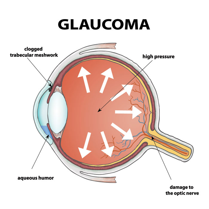
Detecting glaucoma early is critical to preventing permanent vision loss. Regular eye exams play a key role, and these exams often include specialized tests to assess the health of your optic nerve and eye pressure. In this guide, we’ll discuss six common glaucoma tests that eye specialists use to diagnose and monitor the disease.
1. Tonometry
Tonometry measures intraocular pressure (IOP), a major factor in glaucoma development. The most widely used method is Goldmann Applanation Tonometry, considered the gold standard. During the test, anesthetic eye drops are applied, and a device lightly touches the cornea to check pressure levels.
Other tools, like the Tono-Pen and iCare Tonometer, offer portable alternatives, especially useful in clinics and community screenings.
2. Ophthalmoscopy
Ophthalmoscopy helps doctors examine the optic nerve and retina. Using a handheld instrument called an ophthalmoscope, light is directed into the eye to visualize the optic nerve head for signs of damage or structural changes.
This test is vital for determining disease severity and monitoring progression over time.
3. Gonioscopy
The anterior chamber angle, where the iris meets the cornea, plays a critical role in fluid drainage and IOP regulation. Gonioscopy evaluates this angle to identify whether it is open or blocked.
For this test, the doctor uses numbing drops and places a special contact lens called a gonioscope on the eye. This allows clear visualization of the drainage angle and helps guide treatment decisions for open-angle or angle-closure glaucoma.
Planning for treatment? Get the Philippines glaucoma surgery price guide here:
https://glaucoma.ph/glaucoma-treatment-cost-philippines-2025/
4. Visual Field Test (Perimetry)
Peripheral vision loss often occurs early in glaucoma. A visual field test, or perimetry, checks for blind spots in your vision.
You’ll sit in front of a dome-shaped device, focusing on a central target. Small lights flash in different areas, and you indicate when you see them. The results show any gaps in vision, helping the doctor detect early damage and track disease progression.
5. Retinal Nerve Fiber Analysis
Glaucoma damages the retinal nerve fibers, leading to thinning of this layer. Optical Coherence Tomography (OCT) and Scanning Laser Polarimetry (SLP) are advanced imaging techniques that measure this thickness accurately.
These tests create detailed cross-sectional images of the retina, helping identify early nerve damage even before noticeable vision loss.
6. Corneal Thickness Test (Pachymetry)
Corneal thickness affects IOP readings, making pachymetry an important test. A thick cornea can give falsely high pressure readings, while a thin cornea can make pressure appear lower than it is.
The test involves applying numbing drops, then gently touching the cornea with a pachymeter to measure thickness. This data ensures accurate IOP interpretation and guides proper treatment.
Conclusion
These six tests form the foundation of glaucoma detection and monitoring. If you’re at risk or already diagnosed, regular eye exams are essential to protect your vision. Early diagnosis gives you the best chance of preserving sight and preventing irreversible damage.