UK First Surgery Displaces Man’s Eye to Treat Life-Threatening Brain Issue
In a groundbreaking medical achievement, surgeons in the United Kingdom have successfully performed a first-of-its-kind operation involving temporary displacement of a patient’s eye to access and treat a life-threatening brain condition. This rare and highly delicate procedure, carried out at a leading NHS hospital, marks a major milestone in neurosurgery and offers new hope for patients with complex cranial disorders.
The case has drawn significant attention from the global medical community, highlighting the expanding role of innovative surgical techniques in treating conditions once considered inoperable.
A Pioneering Procedure: What Actually Happened?
The surgery, known as a transorbital neuroendoscopic approach, involved carefully repositioning the patient’s eye within the orbit—without removing it or damaging its function—to create a direct surgical corridor to the brain.
Importantly, the eye itself was preserved. The optic nerve and blood supply remained intact throughout the procedure, and the patient retained full vision after surgery.
Why Was This Extreme Step Necessary?
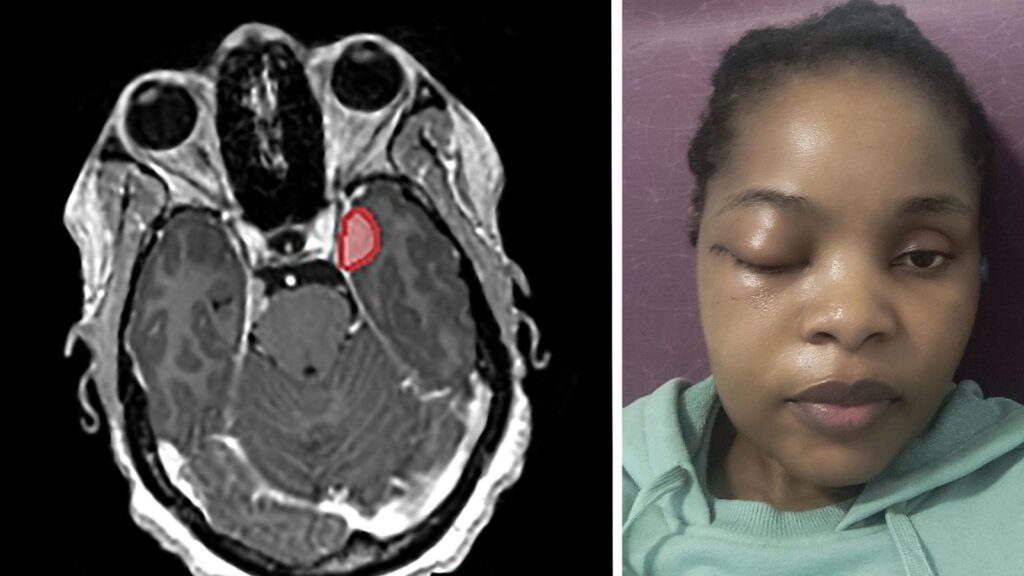
The patient, a man in his 40s, had a large, high-risk brain aneurysm located deep within the skull base near critical regions responsible for vision, speech, and motor control. Its position made traditional surgical approaches extremely risky.
A standard craniotomy carried a high likelihood of severe complications, including bleeding, stroke, or neurological damage. The aneurysm was also pressing against the optic nerve, already causing visual symptoms, and posed an imminent risk of rupture.
An endovascular approach (via catheter through the groin) was also ruled out due to the aneurysm’s complex structure and location.
As a result, the surgical team opted for the transorbital route, allowing direct access through the eye socket while minimizing disruption to surrounding brain tissue and blood vessels.
The Technical Mastery Behind the Operation
This procedure required a highly specialized multidisciplinary team, including neurosurgeons, ophthalmologists, anesthesiologists, and surgical nurses.
Key Steps of the Procedure
- Preoperative Planning: High-resolution MRI and CT scans were used to build a 3D model of the skull, orbit, and aneurysm, enabling precise surgical mapping.
- Orbital Access: A small incision was made in the upper eyelid or conjunctiva, avoiding visible scarring. Orbital structures were carefully exposed.
- Eye Displacement: The eye was gently retracted and stabilized using microsurgical techniques, with continuous monitoring of intraocular pressure to prevent damage.
- Cranial Entry: A high-speed drill was used to create a small opening in the sphenoid bone at the back of the orbit, providing access to the aneurysm.
- Aneurysm Clipping: A titanium clip was placed across the aneurysm to stop blood flow while preserving the parent artery, guided by intraoperative imaging.
- Reconstruction and Closure: The bone window was sealed, the eye was returned to its normal position, and the incision was closed with fine sutures.
Why This Is a “UK First”
Although transorbital approaches have been used internationally for select tumors and sinus-related conditions, applying this technique to a full aneurysm clipping is extremely rare.
The UK team also developed specialized methods for stabilizing the eye during prolonged surgery and maintaining safe surgical conditions within the orbital and sinus regions. In parts of the procedure, the patient was lightly responsive under local anesthesia, allowing real-time monitoring of vision.
Patient Recovery and Outcome
Post-surgery, the patient experienced temporary swelling and mild double vision, which resolved within two weeks. He was discharged after five days and has since returned to normal daily activities.
Follow-up scans confirmed complete aneurysm occlusion, and his vision returned to baseline.
The patient described the experience simply:
“I woke up and thought I had lost my eye, but everything was explained. I felt no pain, and I’m just grateful to be alive.”
Broader Implications for Neurosurgery
This case represents a potential shift in how surgeons approach deep-seated brain conditions.
Advantages of the Transorbital Approach
- Reduced brain manipulation: No need to retract major brain regions
- Faster recovery: Shorter hospital stays compared to open skull surgery
- Better cosmetic outcomes: No visible scalp incisions
- Lower infection risk: Reduced exposure to nasal and sinus pathways
However, experts emphasize that this technique is highly selective. It is only suitable for specific anatomical conditions where safe access through the orbit is possible.
The Future of “Eye-Sparing” Brain Surgery
The success of this UK first opens possibilities for expanding transorbital techniques to other conditions, including:
- Skull base and orbital tumors
- Arteriovenous malformations (AVMs)
- Complex facial and orbital trauma
Researchers are also exploring robotic-assisted tools to improve precision during eye manipulation and orbital access. A multicenter NHS study has been proposed to standardize outcomes and refine surgical protocols.
What Patients Should Know
Patients with complex brain conditions should seek care at specialized neurosurgical centers capable of offering multiple treatment approaches.
Key questions to ask include:
- Is my condition suitable for a transorbital approach?
- What imaging and planning tools are used preoperatively?
- What are the risks compared to traditional surgery?
- Is there a backup plan if this approach is not feasible?
As one lead surgeon noted:
“We didn’t just treat a disease—we preserved quality of life. That is the goal of modern neurosurgery.”
A Milestone in Medical History
The UK’s first operation involving eye displacement for brain treatment represents a significant step forward in surgical innovation. It demonstrates how advances in anatomy, imaging, and microsurgical tools are reshaping what is possible in neurosurgery.
For the patient, it meant avoiding a high-risk skull operation and preserving vision. For medicine, it marks another step in redefining the boundaries between minimally invasive and traditionally “inoperable” brain surgery.