Leeds Surgeons Make UK History with Brain Surgery Through Eye Socket
In a groundbreaking medical achievement, a team of surgeons in Leeds has performed the first-ever brain surgery in the UK conducted entirely through the eye socket. This minimally invasive procedure, which avoids traditional open-skull methods, marks a significant leap forward in neurosurgery and offers new hope for patients requiring delicate brain interventions. Here, we examine the procedure, its implications, and what this means for the future of neurosurgical care.
Revolutionary Approach: Brain Surgery Via the Eye Socket
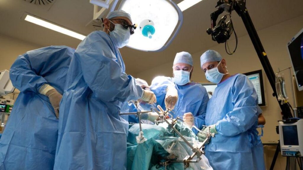
The historic operation took place at Leeds General Infirmary, where a multidisciplinary team successfully removed a brain tumor from a patient using a technique known as transorbital neuroendoscopic surgery (TONES). Instead of making a large incision in the skull, the surgeons accessed the brain through the natural anatomical pathway provided by the bony orbit—the socket that holds the eye.
By carefully navigating instruments through this narrow corridor, they reached the targeted region of the brain without disturbing critical structures such as nerves, blood vessels, or the frontal lobe.
This approach is not entirely new in international neurosurgery, but its successful execution in the UK for a full-scale brain tumor removal is a first. The team, led by consultant neurosurgeon Mr. James Loan, spent months preparing through cadaveric dissections and advanced simulation to ensure precision.
Why This Matters: Benefits Over Traditional Craniotomy
Traditional brain surgery often requires a craniotomy, where a portion of the skull is temporarily removed to expose the brain. While effective, this method carries risks such as infection, prolonged recovery, visible scarring, and potential damage to brain tissue. The transorbital route significantly mitigates these concerns.
Key advantages of transorbital brain surgery include:
- Reduced recovery time: Patients can often leave the hospital within days rather than weeks
- Minimal scarring: The incision is made within the eyelid or behind the eye, leaving virtually no visible mark
- Lower infection risk: By avoiding scalp and skull exposure, infection risk is reduced
- Preservation of brain function: The approach avoids manipulating the frontal lobes
- Shorter anesthesia time: The procedure typically takes less time than a standard craniotomy
For the patient, a 55-year-old woman suffering from seizures and vision disturbances, the outcome was transformative. She was discharged within 48 hours and reported minimal discomfort compared to expectations of open surgery.
The Procedure: How Surgeons Navigated the Eye Socket
The surgery is a showcase of modern technology and anatomical precision. Here is a step-by-step breakdown of how the Leeds team achieved this UK first.
Step 1: Pre-operative Planning
High-resolution MRI and CT scans were fused to create a 3D model of the skull, brain, and tumor location. Neuronavigation software—similar to GPS—was used to map the safest trajectory.
Step 2: Accessing the Orbit
Under general anesthesia, surgeons made a small incision in the upper eyelid crease. The eye was gently retracted to expose the orbital bone. A small window, about the size of a coin, was created in the orbital roof using a micro-drill.
Step 3: Navigating the Brain
A rigid neuroendoscope was inserted through the opening. It provided high-definition visualization of the brain and tumor. Micro-instruments, no wider than a pencil lead, were used to carefully dissect the tumor.
Step 4: Tumor Removal and Closure
The tumor was removed in small pieces while preserving the optic nerve and surrounding vessels. Once complete removal was confirmed using imaging, the area was closed with a titanium plate, and the eyelid incision was sutured.
Patient Impact: A Life Changed
The patient, who wished to remain anonymous, described the experience as “life-changing.” Before surgery, she suffered daily seizures and loss of independence.
Two weeks after the procedure, she had resumed walking, reading, and driving, without the prolonged recovery associated with traditional brain surgery.
“I woke up feeling like I had only had an eye operation, not brain surgery,” she said. “I cannot thank the surgeons enough for giving me back my life with so little disruption.”
The Science Behind Transorbital Neurosurgery
The orbit is a pyramidal cavity surrounded by bone, but its roof is extremely thin—sometimes less than 1 millimeter. This natural anatomical weakness allows access to the anterior cranial fossa.
Historically, this pathway was used in ophthalmology and ENT surgery. Its extension into neurosurgery required mastery of operating in deep, narrow spaces while avoiding critical structures.
Types of Tumors Suitable for This Approach
Not all tumors are suitable. The Leeds team selected a tumor in the anterior skull base region.
Suitable cases include:
- Meningiomas of the olfactory groove or planum sphenoidale
- Pituitary adenomas with anterior extension
- Frontal lobe gliomas in inferior regions
- Cysts or abscesses in the anterior cranial fossa
Deeper brain tumors still require traditional craniotomy.
Challenges and Risks: Not Without Caution
Despite being minimally invasive for patients, the technique is highly complex for surgeons. The narrow operating corridor increases technical difficulty.
Potential risks include:
- Double vision from eye muscle disturbance
- Cerebrospinal fluid leak
- Bleeding in confined spaces
- Infection, though less common than in open surgery
Advanced simulation training and specialized instruments help reduce these risks.
The Future of Neurosurgery in the UK
This procedure reflects a broader shift toward minimally invasive neurosurgery. Similar approaches are being explored for spinal conditions, vascular disorders, and aneurysms.
Mr. James Loan stated:
“This is just the beginning. We are exploring broader applications, including skull base repair and targeted brain therapies.”
Training the Next Generation
The Leeds team is developing structured training programs involving cadaver labs, virtual reality simulation, and supervised procedures.
Healthcare System Impact
The approach may reduce hospital stays, ICU usage, and rehabilitation time, potentially lowering treatment costs for suitable cases within the NHS.
What Patients Should Know
Transorbital surgery is not widely available and applies only to specific tumor locations.
Questions to ask include:
- Is my tumor accessible through the orbit?
- What experience does the surgeon have with minimally invasive techniques?
- What is the recovery time compared to traditional surgery?
- Are there specialist centers offering this procedure?
Conclusion: A New Era in Brain Surgery
Leeds surgeons have demonstrated that brain surgery through the eye socket is both feasible and effective in selected cases. This milestone combines anatomical precision with modern endoscopic technology to reduce patient trauma.
As training expands and techniques evolve, this approach may become an important option in neurosurgical care, reshaping how certain brain conditions are treated.