Optic Nerve Imaging Techniques In Glaucoma Detection
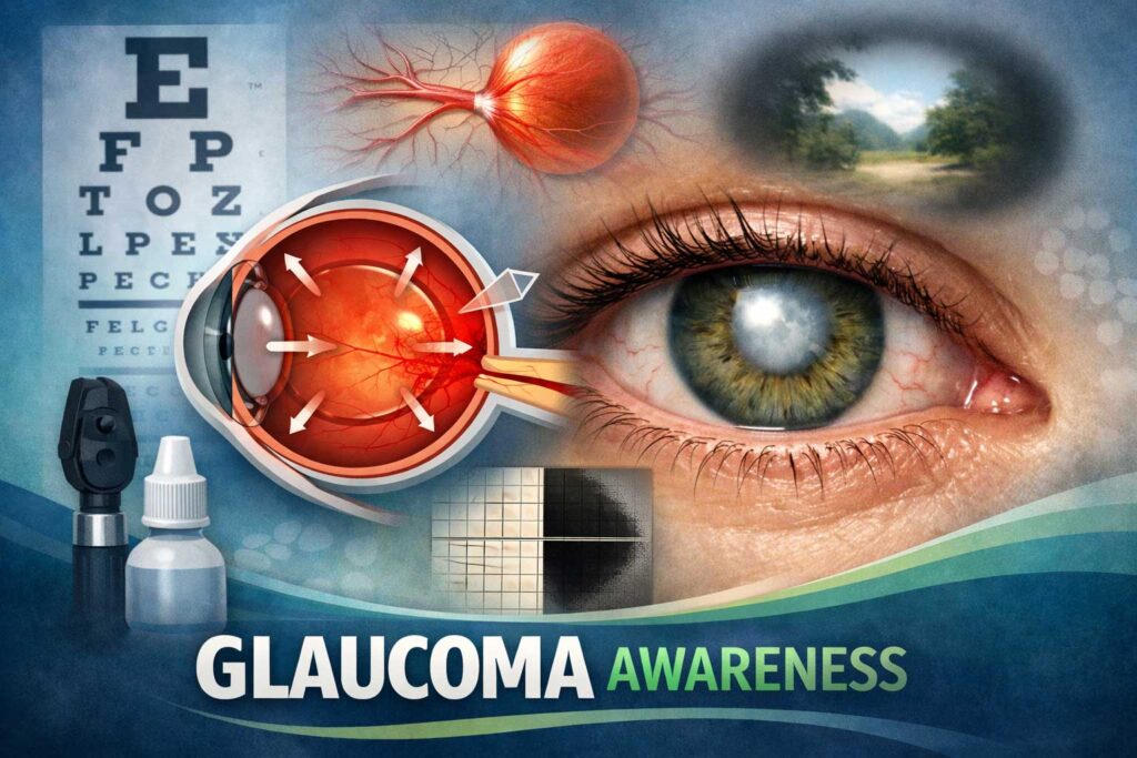
Introduction to Glaucoma and Its Impact
Detecting and managing glaucoma, a leading cause of irreversible blindness worldwide, is crucial for preserving vision. With advancements in technology, optic nerve imaging techniques have revolutionized the way we diagnose and monitor this silent thief of sight. By providing detailed images of the optic nerve, these cutting-edge tools enable early detection and targeted treatment. In this blog post, we will delve into the world of glaucoma detection through optic nerve imaging techniques, exploring their benefits and limitations while uncovering the exciting possibilities that lie ahead. So grab your reading glasses (if you need them!) as we embark on a journey to unravel the secrets behind glaucoma diagnosis!
Overview of Optic Nerve Imaging Techniques
Optic nerve imaging techniques play a crucial role in the detection and management of glaucoma. These advanced technologies provide eye care professionals with valuable insights into the health of the optic nerve, which is essential for diagnosing and monitoring this silent but potentially devastating eye condition.
One widely used technique is Optical Coherence Tomography (OCT), which uses light waves to capture high-resolution images of the retina and optic nerve. It allows for precise measurements of important parameters such as retinal nerve fiber thickness, cup-to-disc ratio, and macular volume. This non-invasive imaging method provides detailed information about structural changes in the optic nerve over time.
Another technique gaining popularity is Heidelberg Retina Tomography (HRT). It utilizes confocal laser scanning technology to create three-dimensional images of the optic disc, enabling accurate assessment of its topography and detecting any signs of damage or progression.
The Glaucoma Diagnosis Variable Corneal Compensation (GDx VCC) is another tool that measures retinal nerve fiber layer thickness using laser polarimetry. By analyzing how light interacts with specific structures in the eye, it can identify early signs of glaucoma before visible damage occurs.
Other emerging technologies like adaptive optics imaging and multi-modal imaging are also being explored to improve our understanding of glaucomatous changes at a microscopic level.
While each technique has its unique strengths and limitations, combining multiple imaging modalities may enhance diagnostic accuracy by providing complementary information about different aspects of glaucoma progression.
In recent years, there have been exciting advancements in incorporating artificial intelligence (AI) into optic nerve imaging analysis. AI algorithms can analyze large datasets more efficiently than manual interpretation alone, aiding clinicians in making faster and more accurate diagnoses. Additionally, AI-powered systems can help detect subtle changes over time that might be missed by human observers.
Looking ahead to the future possibilities with optic nerve imaging for glaucoma detection holds great promise. As technology continues to advance, we can expect further refinement of imaging techniques and the development of
Advancements in Technology for Glaucoma Detection
Advancements in Technology for Glaucoma Detection
Glaucoma, a progressive eye disease that damages the optic nerve, is one of the leading causes of blindness worldwide. Early detection and monitoring are crucial to prevent irreversible vision loss. Fortunately, technological advancements have revolutionized the way we diagnose and manage glaucoma.
In recent years, there has been a surge in cutting-edge imaging techniques specifically designed for glaucoma diagnosis. These technologies offer detailed and precise measurements of the optic nerve structure and function, aiding in early detection and effective management.
One such advancement is Optical Coherence Tomography (OCT), a non-invasive imaging technique that captures cross-sectional images of the retina, optic nerve head, and surrounding tissues. With high-resolution images and quantitative analysis tools provided by OCT devices, ophthalmologists can assess changes in retinal thickness or optic disc parameters over time with great accuracy.
Another remarkable technology is Heidelberg Retina Tomograph (HRT). This scanning laser ophthalmoscopy technique provides three-dimensional images of the optic nerve head using confocal microscopy. It enables clinicians to analyze various parameters like cup-to-disc ratio or neuroretinal rim thickness to detect subtle structural abnormalities associated with glaucoma.
Furthermore, there\’s also GDx Variable Corneal Compensation (VCC) which uses laser polarimetry to measure retinal nerve fiber layer thickness without being affected by corneal birefringence variations. This innovative approach allows for more reliable assessment of glaucomatous damage even in individuals with abnormal corneas or previous surgeries.
These advancements not only provide accurate measurements but also allow for better tracking of disease progression over time. By comparing serial scans obtained from these imaging techniques at different intervals during follow-up visits, doctors can monitor changes in patients\’ ocular health more effectively than relying solely on subjective assessments like visual field testing.
The integration of artificial intelligence (AI) algorithms into optic nerve imaging technology is another promising development. AI can analyze vast amounts of imaging data, identify
Types of Optic Nerve Imaging Techniques: OCT, HRT, GDx VCC, and more
Types of Optic Nerve Imaging Techniques: OCT, HRT, GDx VCC, and more
When it comes to diagnosing glaucoma, optic nerve imaging techniques play a crucial role. These advanced technologies allow ophthalmologists to assess the health of the optic nerve and detect any signs of damage or progression of the disease.
One such technique is Optical Coherence Tomography (OCT), which uses light waves to create detailed cross-sectional images of the retina. This non-invasive procedure provides valuable information about the thickness of the retinal nerve fiber layer and helps in monitoring changes over time.
Another widely used technique is Heidelberg Retina Tomograph (HRT). It utilizes confocal laser scanning technology to capture three-dimensional images of the optic nerve head. By analyzing parameters like cup-to-disc ratio and rim thickness, HRT aids in early detection and management of glaucoma.
Gonioscopy-assisted Transluminal Sclerotomy (GATS) has gained popularity for its ability to visualize deep structures within the eye. It allows for precise identification and localization of pathological features that contribute to increased intraocular pressure – a major risk factor for glaucoma development.
Furthermore, Glaucoma Diagnostics with Variable Cornea Compensation (GDx VCC) measures retinal nerve fiber layer thickness while considering corneal birefringence. This technique provides accurate structural assessment independent from corneal properties.
With advancements in technology continuing at a rapid pace, newer imaging techniques are being developed regularly. For instance, Adaptive Optics Scanning Laser Ophthalmoscopy (AOSLO) enables high-resolution imaging at a cellular level within the retina. This holds immense potential for early detection and personalized treatment strategies.
Each optic nerve imaging technique has its own set of benefits as well as limitations. While OCT offers excellent resolution but may be affected by media opacity or poor fixation during image acquisition; HRT provides precise measurements but may be influenced by factors like disc size and atypical
Benefits and Limitations of Each Technique
Benefits and Limitations of Each Technique:
Optic nerve imaging techniques play a crucial role in the early detection and management of glaucoma. Let\’s take a closer look at some of the commonly used techniques, along with their benefits and limitations.
1. Optical Coherence Tomography (OCT):
OCT provides high-resolution cross-sectional images of the optic nerve head, allowing for precise measurement of retinal thickness. This non-invasive technique enables early detection and monitoring of glaucomatous changes. However, OCT has certain limitations such as its dependence on clear media in the eye and potential artifacts that may affect image quality.
2. Heidelberg Retinal Tomography (HRT):
HRT captures three-dimensional images of the optic nerve using laser scanning technology. It offers excellent reproducibility and can detect subtle structural changes in glaucoma progression. Nevertheless, HRT requires expertise to interpret results accurately, and it may not be suitable for patients with significant media opacities.
3. GDx Variable Corneal Compensation (VCC):
GDx VCC enhances imaging by compensating for corneal birefringence effects which can distort measurements in traditional scanning laser polarimetry methods. This technique provides objective measurements related to nerve fiber layer integrity but is limited by its reliance on corneal compensation algorithms that may not be accurate in certain conditions.
4. Confocal Scanning Laser Ophthalmoscopy:
Confocal scanning laser ophthalmoscopy allows for simultaneous visualization of both structure and function by capturing confocal images while assessing retinal sensitivity through visual field testing or microperimetry.
However, this technique requires pupil dilation which might limit its use in some individuals.
5. Adaptive Optics Imaging:
Adaptive optics imaging utilizes advanced technology to correct aberrations caused by ocular structures, resulting in highly detailed images even at a cellular level.
While promising, it is currently limited to research settings due to its complexity and the need for expensive equipment.
Each optic nerve imaging technique
Integration of Artificial Intelligence in Optic Nerve Imaging
Integration of Artificial Intelligence in Optic Nerve Imaging
Artificial intelligence (AI) has made significant advancements in various fields, and the healthcare sector is no exception. In recent years, AI has been integrated into optic nerve imaging techniques for glaucoma detection, revolutionizing the way this condition is diagnosed and monitored.
One area where AI has shown great promise is in analyzing optic nerve images obtained through optical coherence tomography (OCT). AI algorithms can quickly analyze large volumes of data to detect subtle changes in the structure of the optic nerve head, which may indicate early signs of glaucoma. This technology enables earlier detection and intervention, potentially preventing further vision loss.
Another application of AI in optic nerve imaging is its ability to accurately assess retinal nerve fiber layer thickness using devices like Heidelberg Retina Tomograph (HRT) or scanning laser polarimetry with variable corneal compensation (GDx VCC). By combining machine learning algorithms with these imaging techniques, clinicians can obtain more precise measurements and improve diagnostic accuracy.
Furthermore, AI-powered image processing software allows for automated segmentation and quantification of key features relevant to glaucoma diagnosis. This not only saves time but also reduces human error associated with manual measurement techniques. Additionally, these intelligent systems can generate comprehensive reports that aid ophthalmologists in treatment planning and monitoring disease progression over time.
As research continues to explore the potential of integrating artificial intelligence into optic nerve imaging technologies, we can expect even greater advancements on the horizon. For instance, there are ongoing studies investigating deep learning models trained on large datasets to identify patterns that correlate with different stages of glaucoma.
In conclusion,
The integration of artificial intelligence into optic nerve imaging techniques holds tremendous promise for improving early detection and management strategies for glaucoma. With continued advancements in technology and research efforts focusing on refining AI algorithms specific to glaucoma diagnosis, we are likely to witness significant improvements in patient care outcomes related to this sight-threatening condition.
Future Possibilities with Optic Nerve Imaging for Glaucoma Detection
The field of optic nerve imaging has come a long way in the detection and management of glaucoma. With advancements in technology, researchers and clinicians are constantly exploring new possibilities to further improve this diagnostic tool.
One promising avenue for the future is the integration of artificial intelligence (AI) into optic nerve imaging. AI algorithms have shown great potential in analyzing large amounts of data and identifying patterns that may go unnoticed by human observers. By incorporating AI into optic nerve imaging systems, we can enhance the accuracy and efficiency of glaucoma diagnosis.
Another exciting possibility lies in the development of portable and affordable devices for optic nerve imaging. Currently, most imaging techniques require specialized equipment that is only available in clinical settings. However, with ongoing research and innovation, there is hope for creating compact devices that can be used at home or even on-the-go.
Furthermore, researchers are also looking into multi-modal approaches to optic nerve imaging. This means combining different techniques such as optical coherence tomography (OCT), scanning laser polarimetry (SLP), and confocal scanning laser ophthalmoscopy (CSLO) to obtain a more comprehensive understanding of the structure and function of the optic nerve.
In addition to improving diagnosis, future developments may also focus on monitoring disease progression over time using optic nerve imaging. By regularly capturing images or scans, clinicians can track any changes in the appearance or thickness of the retinal nerve fiber layer – a key indicator of glaucoma progression.
While these possibilities hold great promise for enhancing glaucoma detection and management through optic nerve imaging, it\’s important to remember that continued research and validation are necessary before widespread implementation can occur. Nonetheless, these advancements give us hope for an even brighter future where early detection becomes easier than ever before!
Conclusion
Conclusion
In this ever-evolving world of technology and medicine, optic nerve imaging techniques have revolutionized the detection and management of glaucoma. These advanced imaging tools provide invaluable insights into the structure and health of the optic nerve, allowing for early diagnosis and monitoring of this sight-threatening condition.
From Optical Coherence Tomography (OCT) to Heidelberg Retinal Tomography (HRT), each technique brings its own set of benefits and limitations in glaucoma detection. OCT offers high-resolution cross-sectional images, while HRT provides three-dimensional scans for precise assessment. GDx VCC utilizes polarimetry to measure retinal nerve fiber layer thickness, adding another dimension to our understanding.
The integration of artificial intelligence (AI) further enhances the potential of optic nerve imaging by automating analysis processes and improving diagnostic accuracy. Machine learning algorithms can analyze vast amounts of data with incredible speed, detecting subtle changes that may go unnoticed by human observers.
Looking ahead, there are exciting possibilities on the horizon for optic nerve imaging in glaucoma detection. With ongoing advancements in technology, we can expect even higher precision and faster scanning times. Additionally, AI algorithms will continue to evolve and refine their abilities to detect patterns indicative of glaucomatous damage.
However, it is important to remember that although these imaging techniques play a crucial role in diagnosing glaucoma, they should always be used as part of a comprehensive approach that includes patient history evaluation, intraocular pressure measurement, visual field testing,and clinical examination by an ophthalmologist or optometrist.
In conclusion(Conclusively), optic nerve imaging techniques have transformed how we detect and manage glaucoma. By providing detailed structural information about the eye\’s vital structures like never before,optic nerve imaging aids clinicians in making more informed decisions about treatment plans,maintaining better patient outcomes,and ultimately preserving vision.
Whether it\’s through OCT,HRT,GDx VCC,or future advancements in technology,optic nerve imaging is a game-changer