Cutting-Edge Glaucoma Treatments Unveiled at 2026 ASCRS Meeting
Leading glaucoma experts competed in a dynamic symposium showcasing the latest advances in laser and minimally invasive surgical approaches to treating glaucoma.
A Novel Format for Clinical Education
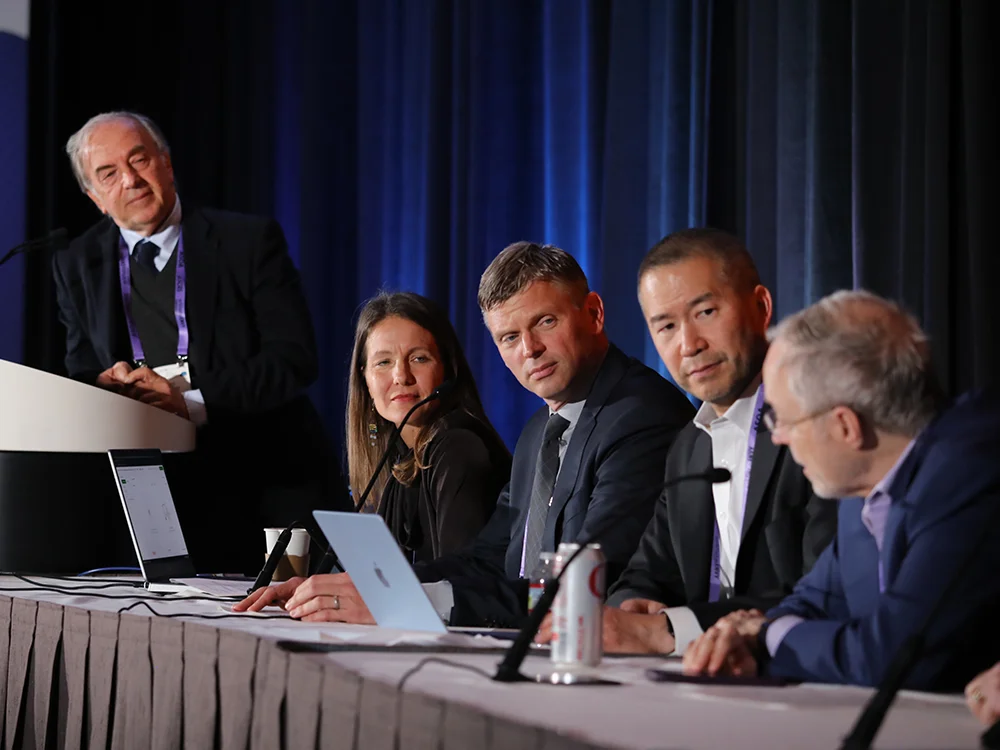
Imagine sitting in a packed auditorium where top glaucoma specialists present breakthrough techniques and case studies—then have the audience vote on whose approach resonates most. That was the experience at the 2026 ASCRS Annual Meeting, where six prominent eye doctors participated in a “Survivor”-themed symposium titled “Glaucoma: Rewind. Refresh. Refine.” Attendees heard presentations from Douglas Rhee, MD, Charles Frank, MD, James Murphy, MD, Ariana Levin, MD, Brian Shafer, MD, and Lorraine Provencher, MD. After each round of presentations, audience members voted to eliminate one speaker, who ceremonially “extinguished their torch” before the next group advanced.
This engaging format allowed clinicians to hear multiple expert perspectives on the same clinical challenges within a single session, creating opportunities to compare and contrast treatment philosophies in real time.
Selective Laser Trabeculoplasty Takes Center Stage
Selective laser trabeculoplasty (SLT) emerged as a dominant theme throughout the laser-focused presentations. The LiGHT Trial and other recent evidence have shifted the standard of care significantly: many doctors now use SLT as first-line therapy for treatment-naïve patients with primary open-angle glaucoma rather than starting with eye drops.
Dr. Rhee highlighted three practical pearls about SLT effectiveness. First, when patients achieve target pressure within two weeks of treatment, there is a 98.2% chance the effect will persist for at least three months—meaning physicians can potentially skip an office visit without compromising outcomes. However, full pressure reduction can take up to six weeks, so early assessment should not be mistaken for final results. Second, a cellular repopulation mechanism occurs at Schwalbe’s line, so practitioners should intentionally space laser shots to allow room for this regeneration process.
Dr. Frank emphasized the importance of post-laser anti-inflammatory medication, noting that the SALT (Steroids After Laser Trabeculoplasty) Trial showed no significant difference between nonsteroidal anti-inflammatory drugs and steroids in terms of pressure-lowering effect—but both were significantly lower than placebo. He advocated for routine anti-inflammatory drops for 5–7 days following any SLT procedure, making it standard care rather than optional.
Dr. Murphy underscored that much about SLT remains unknown: optimal power settings, bubble patterns, distribution zones, and ideal frequency have not been fully characterized. The COAST (Clarifying the Optimal Application of SLT Therapy) Trial is investigating low-power scheduled annual SLT, a protocol that Dr. Murphy has adopted. He stressed that SLT is more cost-effective than topical medications as primary therapy and recommended performing SLT on both eyes whenever possible to reduce administrative overhead.
Dr. Provencher tackled the communication challenge, noting that some physicians hesitate to discuss SLT with patients due to uncertainty about how to frame the conversation. She suggested emphasizing that SLT is first-line, quick, non-invasive, improves aqueous outflow, reduces visual field loss, and decreases the need for future surgeries and drop-related toxicity. Her advice: “stop talking” and let published trial data make the case.
Lasers Beyond Standard Glaucoma
Dr. Levin broadened the discussion to uveitic glaucoma, a more complex form of the disease. She presented multiple case examples showing that lasers can be effective in uveitis-related pressure elevation when combined with a coordinated approach. Her recommendations included collaborating with the uveitis specialist, controlling inflammation beforehand, performing gonioscopy to visualize the angle, treating the underlying cause of elevated pressure, managing inflammation after the laser procedure, and counseling patients that they will likely require additional interventions such as gonioscopy-assisted transluminal trabeculotomy (GATT), tube shunts, or cataract surgery in the future.
Dr. Shafer focused on plateau iris, a particularly challenging condition in which the ciliary body is rotated anteriorly, crowding the angle regardless of how many laser holes are created. He advocated for endoscopic cyclophotocoagulation (ECP) combined with cataract surgery—or performed in an already pseudophakic eye—because this approach can posteriorly rotate the ciliary body and structurally open the angle. This addresses the root anatomic problem rather than simply managing pressure symptoms.
Minimally Invasive Glaucoma Surgery: Multiple Devices, Strategic Selection
The second round of presentations shifted to MIGS (minimally invasive glaucoma surgery), highlighting the growing array of options available to practitioners. Dr. Rhee noted that SLT has limited benefit after failed goniotomy, necessitating a MIGS approach. He discussed comparative effectiveness, explaining that the COMPARE Study demonstrated Hydrus (Alcon) outperforms both two iStents and two iStent inject devices (Glaukos) when more substantial outflow and pressure control are needed. These findings aligned with predictions from preclinical laboratory models.
Dr. Frank addressed spatial constraints during angle surgery: when the nasal angle is occupied and additional angle work is needed, surgeons can access other portions of the angle using mirrored goniolenses and performing goniosynechialysis—adhesion release—to expand treatment options.
Dr. Murphy advocated for immediate sequential bilateral cataract and MIGS procedures, performing goniotomy, canaloplasty, microstent placement, and other angle interventions on the same day. He emphasized hands-free techniques using goniolenses without handles to streamline workflow.
Dr. Shafer presented a nuanced perspective: “nurture the drain, but don’t alter it.” He outlined four MIGS mechanisms—trabecular meshwork bypass stents, goniotomy or trabeculotomy, canaloplasty, and viscodilation—and noted that no single device is universally superior. He favored techniques that preserve the native drainage system because leaving the anatomy intact allows future options if pressure control needs adjustment.
Dr. Provencher described a cyclopexy repair technique for nasal cyclodialysis clefts that can occur during MIGS. She emphasized that all surgeons performing MIGS should understand how to close such clefts when they occur, using a long-curved cannula with 10-0 Prolene passed directly through sclera to reposition the ciliary body.
When to Consult a Glaucoma Specialist
If you have been diagnosed with glaucoma, notice progressive vision loss, experience elevated eye pressure readings, or have a family history of glaucoma, scheduling an appointment with a glaucoma specialist is important. Early discussion about treatment options—whether laser, MIGS, or medications—allows you to understand the full range of approaches and make informed decisions aligned with your long-term eye health goals.
If you undergo SLT, follow-up examination at two weeks helps determine whether target pressure has been reached and whether additional treatment is needed. Those with uveitic glaucoma or plateau iris should work closely with specialists experienced in these conditions, as treatment strategies differ substantially from routine cases.
The Evolving Landscape of Glaucoma Care
The 2026 ASCRS symposium demonstrated that glaucoma management has evolved significantly. Laser therapy is no longer a backup option but a primary intervention with strong evidence supporting its use. MIGS devices continue to expand in number and sophistication, offering surgeons multiple ways to improve aqueous drainage while preserving anatomy. Equally important, the specialty is moving toward individualized treatment selection based on disease subtype, anatomy, and patient preferences rather than applying a one-size-fits-all algorithm.
Staying informed about these advances helps patients ask the right questions during their appointments and understand why their doctor may recommend a particular approach. As more glaucoma specialists adopt these evidence-based techniques, access to cutting-edge care continues to spread beyond specialized centers into community practices.