Parapapillary Atrophy Changes Signal Risk in Angle-Closure
Beta parapapillary atrophy enlargement may precede clinical glaucoma progression in susceptible patients over five years.
parapapillary atrophy glaucoma
What Is Parapapillary Atrophy and Why Does It Matter?
The area surrounding your optic nerve head—the spot where nerves leave your eye—can undergo subtle structural changes that ophthalmologists track carefully. Beta zone parapapillary atrophy (β-PPA) refers to a specific pattern of tissue thinning in this region. While researchers have long recognized a connection between β-PPA and primary open-angle glaucoma, its role in primary angle-closure disease has remained less well understood until recently.
Understanding these changes matters because they may signal early warning signs before glaucoma becomes clinically detectable. A new longitudinal cohort study published in Ophthalmic Research in August 2026 sheds light on how β-PPA evolves in primary angle-closure suspect (PACS) patients—people who have anatomical features suggesting they’re at risk for angle-closure glaucoma but haven’t yet developed the full disease.
The Five-Year Study: What Researchers Found
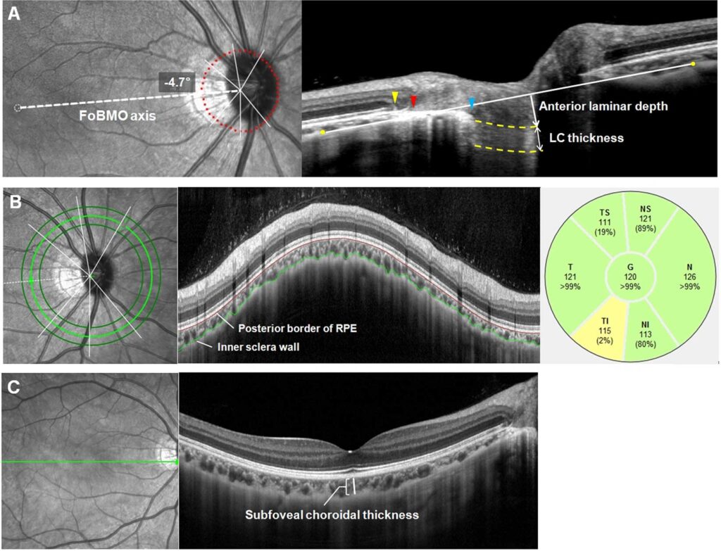
Investigators in China tracked 83 eyes from 83 PACS participants (mean age 61.1) over a five-year period to examine changes in parapapillary atrophy and associated risk factors. Parapapillary atrophy was assessed using stereoscopic fundus photography and verified with optical coherence tomography (OCT), providing both photographic and imaging documentation of any structural changes.
The headline finding: β-PPA enlargement was observed in nearly 27% of PACS eyes over the five-year follow-up period. This enlargement was strongly linked to baseline measurements showing a longer axial length (the front-to-back length of the eye) and a larger vertical cup-to-disc ratio (the proportion of the optic cup to the overall optic disc). These associations suggest that biomechanical or structural remodeling may occur in the eye before clinically detectable glaucomatous optic nerve damage appears.
Understanding Axial Length and Biomechanical Strain
One striking finding involved the role of axial length. Most participants in the study were nonmyopic with relatively short axial lengths—yet longer axial length at baseline correlated with enlargement in three specific measurements: the area, angular extent, and maximum radial length of β-PPA. This discovery challenges a common assumption that biomechanical strain from axial elongation occurs mainly in myopic eyes.
The researchers noted that eyes with shallow anterior chambers, thick lenses, and short axial lengths can display myopic-like features due to accelerated elongation patterns. Even in nonmyopic or relatively short-axial-length eyes, individual differences in axial length or axial elongation-related remodeling may contribute to the peripapillary structural changes observed in the study. This insight broadens our understanding of how mechanical forces shape the optic nerve head region across different eye types.
The Vertical Cup-to-Disc Ratio Connection
The vertical cup-to-disc ratio—a standard measurement used in every eye exam—also emerged as a factor associated with β-PPA enlargement. Although the absolute magnitude of change in this ratio was very small, the study authors suggested that this connection points toward early glaucoma-related structural modifications occurring before any clinical signs of glaucomatous optic neuropathy become visible.
This distinction is important. The changes were present but subtle, which is why only ophthalmologists and optometrists using specialized imaging and measurement techniques can reliably detect them. Your eye care provider compares measurements over time to spot these trends.
Why These Findings Matter for People at Risk
For primary angle-closure suspects—individuals with risk factors like narrow anterior chambers but without frank angle closure or elevated eye pressure—this research adds a new lens to monitoring. If β-PPA enlargement represents an early structural marker of disease progression, it could help identify which suspects are most likely to advance to primary angle closure (PAC) or primary angle-closure glaucoma (PACG).
However, the authors emphasized an important caution. Longer axial length should be considered a potential confounding factor, meaning β-PPA enlargement shouldn’t automatically be interpreted as disease-related progression. Instead, it may reflect a mix of biomechanical remodeling and degenerative changes influenced by individual eye anatomy.
What This Means for Monitoring and Follow-Up
The research highlights why consistent, long-term monitoring matters for PACS patients. Rather than viewing each eye exam as a standalone snapshot, your ophthalmologist or optometrist builds a timeline of measurements. Serial photography, OCT imaging, and standardized cup-to-disc ratio measurements create a record that can reveal slow-progressing changes invisible in a single visit.
The study examined changes over five years, a timeframe that allowed subtle shifts to become measurable. This underscores why people with angle-closure risk factors need scheduled follow-up appointments rather than sporadic visits—the changes are gradual and only become meaningful when compared across time.
The Bigger Picture: Where Research Goes From Here
The authors acknowledged that while their findings are suggestive, they raise more questions than they answer. They called for future prospective cohort studies with longer follow-up periods to determine whether β-PPA enlargement can reliably serve as an early structural indicator or predictor of progression along the primary angle-closure disease spectrum.
Translation: this study is an important first step, but researchers need to track larger groups of PACS patients over even longer periods to confirm whether β-PPA changes consistently predict who will progress to more advanced angle-closure disease. That kind of evidence would eventually help guide clinical decision-making about who needs preventive treatment and when.
When to Schedule an Eye Exam
If you have been told you are a primary angle-closure suspect, or if you have a family history of angle-closure glaucoma, don’t skip regular appointments. Schedule a comprehensive eye exam if you have never been screened for angle-closure risk, especially if you have risk factors like hyperopia (farsightedness), a shallow anterior chamber, or a thick lens relative to your eye size.
Seek urgent care if you experience sudden eye pain, blurred vision, halos around lights, eye redness, or nausea—these can signal an acute angle-closure episode and require immediate treatment to prevent permanent vision loss.
Between appointments, monitor your vision for gradual changes in clarity or visual field. While most PACS patients never progress to acute angle closure, consistent monitoring gives your eye care team the best chance to detect early structural changes and intervene if needed. The five-year study findings suggest that seemingly minor anatomical differences—axial length, cup-to-disc ratio, and now β-PPA changes—may collectively paint a picture of future risk, making continuity of care and serial measurements invaluable tools for vision preservation.