Lab-Grown Retina Reveals Genetic Cause of Rare Childhood Eye Disease
For families navigating the difficult diagnosis of a rare childhood eye disease, the path forward is often shrouded in uncertainty. Many conditions lack clear genetic explanations, making prognosis and treatment planning a formidable challenge. Now, a groundbreaking study has illuminated one such shadowy corner of pediatric ophthalmology, not in a living patient, but within the intricate layers of a tiny, lab-grown retina.
A Miniature Breakthrough in a Dish
Researchers have successfully used retinal organoids—three-dimensional, miniature replicas of the human retina grown from stem cells—to pinpoint a previously unknown genetic culprit behind a severe form of Leber Congenital Amaurosis (LCA). LCA is a group of disorders that causes severe vision loss or blindness from infancy. While many genes have been linked to LCA, a significant number of cases remain genetically unsolved.
This research, a powerful fusion of stem cell technology and genetic detective work, offers more than just an answer. It provides a revolutionary model for understanding how specific genetic errors literally dismantle the eye’s essential light-sensing machinery, cell by cell.
From Patient Cells to a Living Laboratory
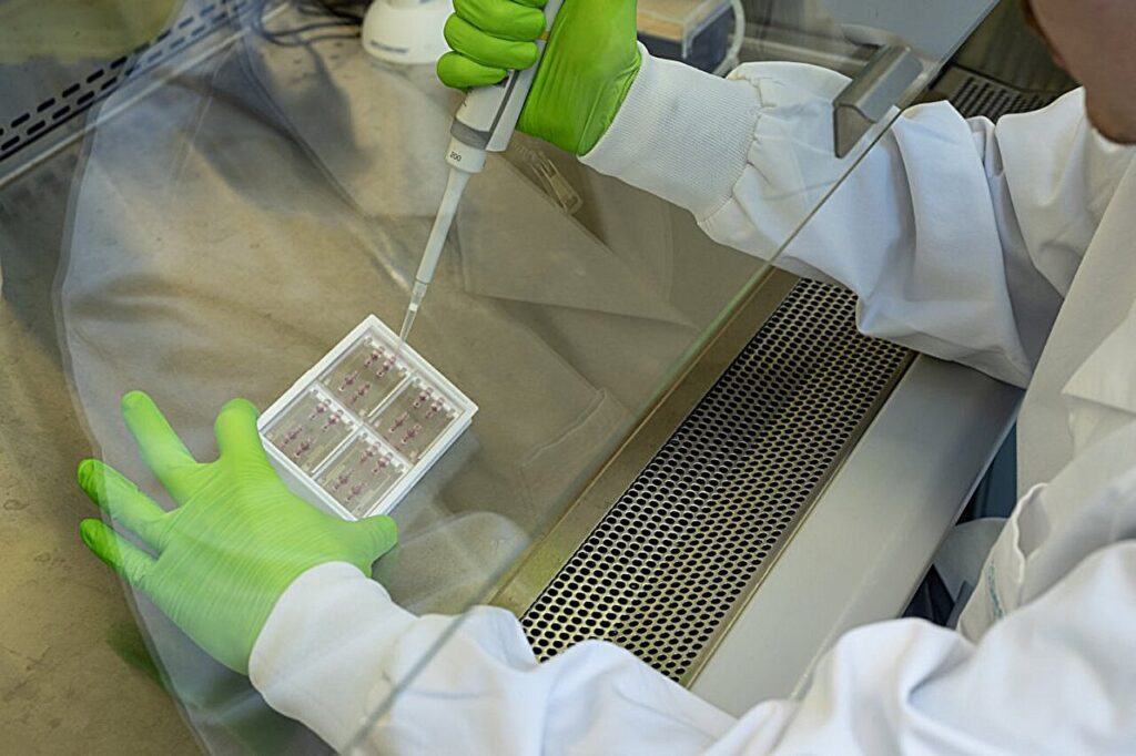
The journey began with skin cells taken from a young patient with a severe, yet genetically undiagnosed, form of LCA. Scientists reprogrammed these skin cells into induced pluripotent stem cells (iPSCs), a type of cell with the remarkable potential to become almost any other cell in the body.
The Power of the Organoid
From these iPSCs, the team meticulously grew retinal organoids. Over several months, these clusters of cells self-organized into the complex, layered structure of a human retina, complete with developing photoreceptors—the rods and cones that convert light into electrical signals for the brain.
Crucially, the patient-derived organoids showed clear and devastating defects. The photoreceptors, particularly the cone cells responsible for color and central vision, failed to develop properly. Their light-sensing outer segments, critical for vision, were stunted or completely absent. The researchers now had a perfect living laboratory to trace this physical failure back to its genetic source.
The Genetic Culprit Unmasked
Using advanced whole-genome sequencing on the organoids, the research team hunted for the error. They discovered a mutation in a gene that had not been previously associated with LCA. This gene is responsible for producing a protein essential for the maintenance and function of the primary cilium.
Why the Cilium is Key
This finding was a revelation. The photoreceptor’s light-sensing outer segment is a specialized type of primary cilium. Think of the cilium as a cellular antenna; in photoreceptors, this antenna is expanded and packed with light-sensitive molecules.
- The Mutation: The newly identified mutation disrupts the structure of this critical protein.
- The Breakdown: The faulty protein cannot support the cilium, causing it to collapse during its crucial development phase.
- The Result: Without a stable cilium, the photoreceptor cannot build its light-sensing outer segment. The cell is left functionally blind, leading to the profound vision loss seen in the patient.
“This was a classic ‘chicken and egg’ problem in genetics,” explained a senior researcher on the project. “We could see the photoreceptors were dying, but we didn’t know the initial molecular event that doomed them. The organoid model let us watch the disease unfold from its very beginning and identify the precise point of failure.”
Beyond a Single Diagnosis: A New Pathway for Discovery
The implications of this study stretch far beyond diagnosing one form of LCA. It validates a powerful new paradigm for researching inherited retinal diseases (IRDs).
- Solving Unsolved Cases: For the estimated 20-30% of IRD patients without a genetic diagnosis, organoid technology provides a dynamic tool to find answers where standard genetic testing has failed.
- Understanding Disease Mechanisms: Scientists can now observe the real-time consequences of genetic errors in human tissue, moving from a static DNA report to a moving picture of cellular catastrophe.
- Testing Therapies: These patient-specific organoids become the ultimate testbed for potential therapies. Researchers can use them to trial gene correction techniques (like CRISPR), drug compounds, or neuroprotective agents directly on the affected human retinal cells before ever moving to a clinical trial.
A Future Forged in the Lab
The story of this discovery is a testament to how modern biology is converging to tackle rare diseases. It blends patient-derived stem cells, sophisticated 3D tissue culture, and cutting-edge genomics to create answers—and hope—where none existed.
For the family of the child at the heart of this study, the discovery provides a long-sought explanation. For the scientific and medical community, it opens a direct window into the earliest stages of blinding diseases. Perhaps most importantly, it establishes a reproducible path forward: grow, observe, identify, and test.
As retinal organoid technology continues to mature, it promises to demystify more of ophthalmology’s darkest genetic puzzles, guiding the way toward future treatments that could preserve or restore the precious gift of sight for children around the world. The light at the end of the tunnel for rare eye diseases may very well be generated in a lab dish.