What Is the Trabecular Meshwork and Its Role in Glaucoma
A tiny structure inside your eye may hold the key to preventing one of the world’s leading causes of blindness.
You have probably never thought about the trabecular meshwork. Most people have not. It is a microscopic structure buried deep inside the eye, invisible without specialized equipment, and rarely mentioned outside of a doctor’s office. But in 2026, this tiny piece of eye anatomy has become one of the most talked-about topics in medical research — and for very good reason.
Scientists are making extraordinary breakthroughs in understanding how the trabecular meshwork works, what happens when it breaks down, and how new treatments ranging from vitamin supplements to stem cell therapy could revolutionize the way we treat glaucoma. If you care about your vision — or the vision of someone you love — this is a topic worth understanding.
What Is the Trabecular Meshwork?
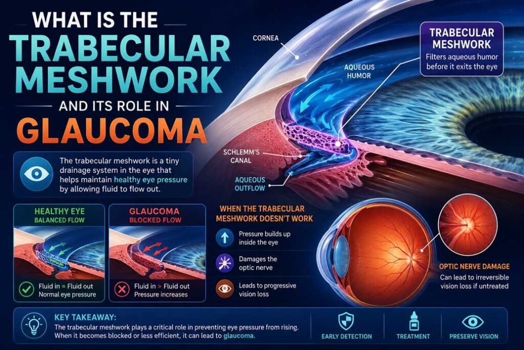
The trabecular meshwork is a small, spongy, ring-shaped tissue located at the drainage angle of the eye — the point where the iris meets the base of the cornea. Despite its tiny size, it performs one of the most critical jobs in the entire eye: managing fluid drainage.
Your eye constantly produces a clear fluid called aqueous humor. This fluid nourishes the lens and cornea, maintains the shape of the eye, and keeps internal pressure at a healthy level. After circulating through the eye, this fluid needs to drain out. The trabecular meshwork acts as the eye’s primary drainage filter, allowing fluid to pass through its porous tissue into a channel called Schlemm’s canal, and from there into the bloodstream.
When this system works properly, eye pressure stays balanced and vision remains healthy. When the trabecular meshwork becomes damaged, stiff, or clogged, fluid builds up, pressure rises, and the optic nerve begins to deteriorate. That deterioration is glaucoma.
The Link Between the Trabecular Meshwork and Glaucoma
Glaucoma is one of the leading causes of permanent, irreversible blindness worldwide. It affects tens of millions of people globally and is particularly dangerous because it progresses silently. Most people do not realize they have it until significant vision loss has already occurred.
The most common form, primary open-angle glaucoma, is directly tied to trabecular meshwork dysfunction. As the meshwork loses cells, stiffens, and becomes less efficient at filtering fluid, outflow resistance increases. Pressure builds inside the eye. Over time, that elevated pressure damages the optic nerve, causing permanent blind spots that gradually expand.
What makes this especially concerning is that the trabecular meshwork naturally loses cells as we age. Combined with genetic factors, inflammation, steroid use, and other contributors, this age-related decline can tip the balance toward glaucoma in many people — often without any warning signs whatsoever.
A Landmark 2026 Discovery: Cell Subtypes and Vitamin B3
One of the most exciting scientific developments this year has completely changed how researchers understand the trabecular meshwork at the cellular level.
Using a cutting-edge technique called single-cell epigenomic profiling, a team of researchers mapped the cellular makeup of the trabecular meshwork in unprecedented detail. What they found surprised everyone: the trabecular meshwork is not a uniform tissue. It contains three distinct cell subtypes, each with its own unique function and genetic signature.
The first subtype, TM1, is primarily responsible for maintaining and remodeling the extracellular matrix — the structural scaffolding that holds the tissue together. The second subtype, TM2, sends signaling molecules to support the neighboring Schlemm’s canal cells. The third subtype, TM3, is the most metabolically active and relies heavily on mitochondrial function to do its job.
It is TM3 that turned out to be the key to understanding glaucoma development. In mice carrying a genetic mutation linked to high eye pressure, TM3 cells showed dramatic signs of mitochondrial dysfunction. Their mitochondria were swollen and structurally damaged — unable to produce the energy the cells needed to function properly. As a result, eye pressure rose.
The truly remarkable finding came next. When researchers treated these mice with vitamin B3 — also known as nicotinamide — the mitochondrial damage was significantly reduced, and eye pressure was protected from rising.
Vitamin B3 is a widely available, affordable, and well-tolerated supplement already used for various health purposes. The possibility that it could play a role in protecting the trabecular meshwork from the kind of cellular damage that initiates glaucoma is a finding that has sent ripples through the ophthalmology world. Human clinical trials exploring this connection are now being closely watched.
Could Stem Cells Regenerate the Trabecular Meshwork?
The vitamin B3 discovery is remarkable, but it is just one part of a much larger scientific revolution taking place around the trabecular meshwork. Researchers are now actively exploring whether the damaged tissue can be physically rebuilt using stem cell therapy.
The concept is straightforward: if glaucoma is caused in part by the loss of trabecular meshwork cells, then replenishing those cells with stem cells could restore normal fluid drainage and bring eye pressure back under control.
Several types of stem cells have been investigated for this purpose, including tissue-specific stem cells, induced pluripotent stem cells derived from a patient’s own body, and adult mesenchymal stem cells. Studies in animal models and laboratory settings have already shown that these stem cells can successfully integrate into the trabecular meshwork, restore cell populations, and improve fluid outflow.
The potential here is enormous. Rather than simply managing glaucoma with lifelong eye drops or repeated surgeries, stem cell therapy could one day address the root cause of the disease — actually repairing the damaged drainage tissue that started the problem in the first place. Early clinical signals are encouraging, and researchers working on this approach have described it as potentially reshaping long-term expectations for glaucoma patients entirely.
Surgical Advances: MIGS and the New Laser Devices
While stem cell therapy is still working its way through early clinical development, there are already meaningful surgical options available today that directly target the trabecular meshwork.
Micro-invasive glaucoma surgery, commonly known as MIGS, has become one of the fastest-growing areas of ophthalmic surgery. Trabecular meshwork-based MIGS procedures work by removing, bypassing, or opening up the obstructed drainage tissue to allow fluid to flow more freely. Compared to traditional glaucoma surgeries, MIGS procedures offer a higher safety profile, better preservation of normal eye anatomy, faster recovery times, and a reduced need for ongoing pressure-lowering medications.
On the laser front, a new generation of selective laser trabeculoplasty devices is making the procedure more accessible than ever before. Traditional laser trabeculoplasty required significant surgical skill, a specialized contact lens, and precise visualization of the trabecular meshwork. A new direct SLT device changes all of that — the surgeon simply points the device at the eye, and within seconds it automatically locates the trabecular meshwork and delivers 100 precisely targeted laser spots without any contact lens required. This dramatically lowers the barrier to performing the procedure and opens it up to a wider range of clinical settings.
New Drug Treatments in the Pipeline
The pharmaceutical industry is also intensely focused on the trabecular meshwork as a treatment target. Several new classes of drugs are in advanced development or already reaching the market.
Rho kinase inhibitors work by relaxing the cellular structure of the trabecular meshwork, making it more pliable and improving fluid outflow. Nitric oxide donors similarly relax the meshwork tissue and have shown strong results in clinical trials. These represent a new generation of glaucoma medications that work differently from traditional eye drops, which typically focused on reducing fluid production rather than improving drainage.
Newer compounds are also targeting the blood vessels and channels downstream of the trabecular meshwork, reducing the resistance that builds up in the broader outflow system. This multi-pronged pharmaceutical approach reflects a growing understanding that glaucoma management requires targeting the entire drainage pathway, not just one point of resistance.
Why Is This Trending Right Now?
The surge of interest in the trabecular meshwork in 2026 is not a coincidence. It is the result of a remarkable convergence of scientific breakthroughs arriving at the same time — the vitamin B3 cell subtype discovery, advancing stem cell research, new surgical devices, and a robust drug pipeline all landing together and painting a picture of a field on the verge of transformation.
For decades, glaucoma treatment was about damage control. Today, the conversation has shifted toward prevention and restoration. That is a fundamental change in direction, and it is generating enormous excitement among both researchers and patients.
What You Should Do Now
If you are over 40, have a family history of glaucoma, or have been told your eye pressure is on the high side, do not wait. Schedule a comprehensive eye exam with an ophthalmologist. The trabecular meshwork cannot be examined without specialized clinical tools — but those tools exist, and early detection remains the most powerful weapon against vision loss.
The science is advancing rapidly. But none of it can help you if the damage is caught too late.
Disclaimer: This article is for informational purposes only and does not constitute medical advice. Please consult a qualified eye care professional for diagnosis and treatment.