AI in Glaucoma Care: Transforming Detection and Patient Communication
Artificial intelligence is revolutionizing how eye doctors detect, monitor, and discuss glaucoma with their patients, offering earlier interventions and more personalized care conversations.
AI glaucoma detection and patient communication
How Artificial Intelligence is Changing Glaucoma Detection
Glaucoma affects millions of people worldwide, yet many don’t realize they have it until significant vision loss has already occurred. The condition develops when intraocular pressure damages the optic nerve, often without causing noticeable symptoms in its early stages. Traditional screening methods rely heavily on manual assessment and clinical judgment, which can sometimes miss subtle signs of disease progression.
Artificial intelligence is reshaping this landscape by analyzing medical imaging with unprecedented precision. AI algorithms trained on thousands of retinal scans and visual field tests can now identify patterns that might escape even experienced clinicians’ eyes. These systems evaluate optical coherence tomography (OCT) images, fundus photographs, and other diagnostic data to detect thinning of the retinal nerve fiber layer—a hallmark of glaucomatous damage.
What makes this particularly valuable is speed and consistency. An AI system can process imaging results in seconds, flagging patients who may benefit from closer monitoring or more aggressive treatment. This means ophthalmologists have more time to focus on complex cases and patient counseling rather than spending hours reviewing routine images.
Earlier Detection Means Better Long-Term Outcomes
The window for preventing glaucoma-related vision loss is critical. Once optic nerve damage occurs, it’s permanent—but catching the disease early allows doctors to slow or halt its progression through various interventions. Intraocular pressure management remains the primary treatment strategy, whether through medications, laser procedures, or surgery.
AI-assisted screening makes it possible to identify at-risk patients before they experience measurable vision loss. Patients with elevated intraocular pressure readings during tonometry, a family history of glaucoma, or other risk factors can receive targeted monitoring protocols. The technology particularly helps in under-resourced settings where specialist eye care may be limited, democratizing access to high-quality glaucoma screening.
Research presented at conferences like ESCRS 2026 highlights how these AI systems reduce false negatives—cases where glaucoma is present but goes undetected. When combined with traditional clinical assessment, AI acts as a safety net, catching disease progression that might otherwise be overlooked during routine exams.
Transforming How Doctors Talk to Patients About Glaucoma
Understanding a glaucoma diagnosis can be overwhelming for patients. The condition is often asymptomatic, raising questions about why treatment is necessary if they feel fine. Doctors must balance urgency with reassurance, explaining complex concepts like intraocular pressure and optic nerve damage in ways that resonate with individual patients.
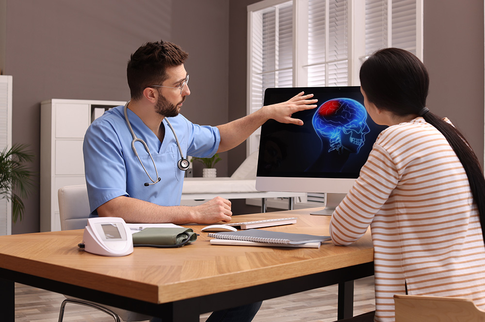
AI is now enhancing these conversations by providing visual evidence of disease progression. Instead of pointing to abstract numbers on a printout, physicians can show patients side-by-side comparisons of their retinal imaging over time, with AI-highlighted areas indicating where thinning has occurred. This concrete visualization helps patients grasp why consistent eye pressure management matters.
Personalized risk stratification powered by AI also allows doctors to tailor their conversations to each patient’s specific situation. Someone with a strong family history, advanced age, and elevated pressure receives a different counseling approach than a younger patient with borderline readings and no family risk. AI can calculate individualized glaucoma progression risk over the next five or ten years, giving patients a clearer picture of their stakes.
The Role of AI in Monitoring Treatment Response
Patients on glaucoma therapy need regular follow-up to ensure their intraocular pressure is adequately controlled and that optic nerve damage isn’t advancing. These visits traditionally involve repeating visual field tests, OCT imaging, and other assessments—some of which can be uncomfortable or time-consuming.
AI algorithms can now predict how quickly a patient’s glaucoma might progress based on their baseline characteristics and early response to treatment. This allows doctors to identify patients who aren’t responding adequately to their current medication regimen and need escalation to combination therapy or laser treatment. Rather than waiting six or twelve months to notice lack of improvement, AI analysis can flag these cases sooner.
Machine learning models also help distinguish true progression from normal measurement variability. Visual field tests and OCT measurements naturally fluctuate slightly from visit to visit. AI can filter out this noise to identify genuine worsening, preventing unnecessary treatment changes while catching real problems early.
Addressing Accuracy and Clinical Integration
While AI shows tremendous promise, its integration into clinical practice requires careful validation and integration with physician expertise. These systems are tools to augment clinical judgment, not replace it. An ophthalmologist’s assessment of a patient’s overall health, medication history, and individual circumstances remains essential to decision-making.
AI systems must also be validated across diverse patient populations to ensure they perform accurately in people with different skin tones, ethnicities, and genetic backgrounds. Early iterations of medical AI have sometimes shown performance gaps in underrepresented groups, highlighting the importance of inclusive training data.
At major conferences like ESCRS 2026, eye care specialists discuss how to implement these technologies responsibly, establishing clear protocols for when AI recommendations should prompt clinical action versus when they serve as supportive information. The goal is seamless integration that enhances workflow without creating alert fatigue or overwhelming clinicians with false concerns.
Patient Communication Benefits Beyond Detection
AI doesn’t just help doctors identify glaucoma—it also supports patient education and compliance. Patients who understand their disease progression and see visual evidence of why treatment matters are more likely to use their eye drops consistently and attend follow-up appointments.
Some AI systems generate automated reports that explain results in patient-friendly language, describing what their intraocular pressure means and how their optic nerve appearance compares to previous visits. This supplementary material helps patients remember key points from their appointment and reinforces the importance of adherence to their prescribed regimen.
Interactive tools powered by AI can also help patients understand their risk factors and lifestyle modifications that might support eye health, such as the importance of regular exercise, blood pressure management, and medication compliance—all factors that may influence glaucoma progression.
When to Schedule Your Eye Examination
If you’re over 40, have a family history of glaucoma, take corticosteroid medications, or have elevated intraocular pressure, you should have comprehensive eye exams at least every one to two years. Symptoms like eye pain, blurred vision, or halos around lights warrant urgent evaluation.
Even without risk factors, adults should receive baseline glaucoma screening around age 40. Early detection through routine exams—potentially enhanced by AI-assisted analysis—remains the most effective way to prevent vision loss from this silent disease.
Schedule an appointment with an ophthalmologist to establish your baseline eye health status and discuss whether more frequent monitoring is appropriate for your individual risk profile. With AI now supporting clinical care, there’s never been a better time to prioritize glaucoma screening and protection of your vision.