How the Eye’s Drainage System May Help Explain Glaucoma
Glaucoma can gradually damage the optic nerve, sometimes before a person notices changes in their vision. One important factor associated with the disease is increased resistance to aqueous humor drainage, which can contribute to elevated pressure inside the eye.
Scientists are investigating how the microscopic structures inside eye cells influence this drainage process. A research study published through Nature examined how microtubule stability affects Schlemm’s canal cells, their mechanical behaviour and the movement of fluid through the eye.
The findings connect the internal structure of these cells with their ability to form drainage pores and regulate aqueous humor outflow. The experiments also identified differences between normal and glaucoma-derived Schlemm’s canal cells.
What Is Schlemm’s Canal?
Schlemm’s canal is a circular, vessel-like channel located near the outer edge of the eye’s anterior chamber. It forms an important part of the conventional aqueous humor drainage pathway.
Aqueous humor is the clear fluid that continuously circulates inside the eye. It supplies nutrients to structures such as the cornea and lens before passing through the trabecular meshwork and entering Schlemm’s canal.
From there, the fluid moves into collector channels and eventually reaches the bloodstream. This drainage process helps maintain normal intraocular pressure.
When resistance along this pathway increases, fluid may not leave the eye as efficiently. Elevated intraocular pressure is an important risk factor for glaucoma-related optic nerve damage.
What Are Microtubules and Why Do They Matter?
Microtubules are microscopic structures inside cells that form part of the cytoskeleton. They help maintain cell shape, organise internal components and support the movement of materials within cells.
They also influence how cells respond to physical forces. Their stability can affect cellular stiffness, internal signalling and the ability of cells to contract.
Schlemm’s canal cells are exposed to mechanical forces created by fluid movement and pressure differences. Their ability to respond to these forces is important for maintaining the drainage pathway.
The researchers investigated whether changing microtubule stability could influence these cellular properties and improve the formation of openings that allow aqueous humor to pass through the canal wall.
Microtubule Stability Affects Cell Contractility
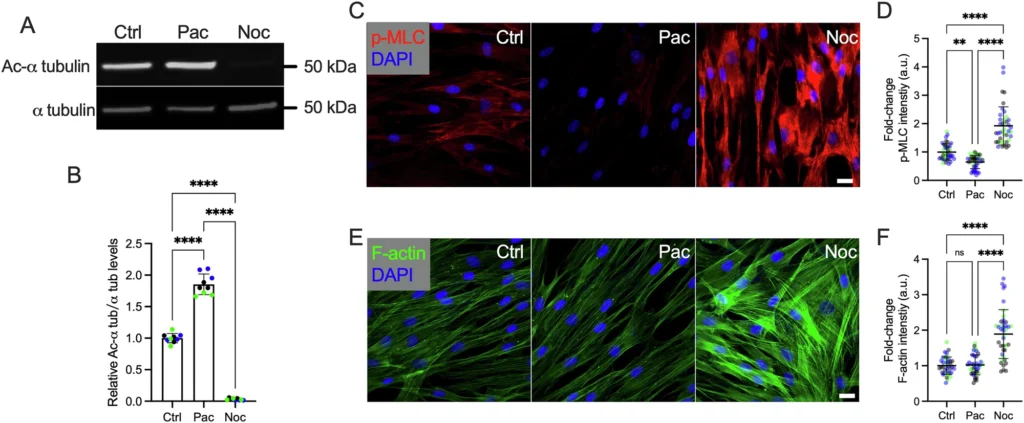
The first experiment examined how stabilising or destabilising microtubules affected actomyosin contractility in normal Schlemm’s canal cells.
Actomyosin is a cellular system involving actin filaments and myosin proteins. It helps cells generate contractile forces and influences their mechanical behaviour.
The researchers used paclitaxel to stabilise microtubules and nocodazole to destabilise them. Their experiments showed that microtubule stabilisation reduced contractility, while destabilisation increased it.
Measurements of phosphorylated myosin light chain (p-MLC) and F-actin helped demonstrate changes in the cells’ contractile activity and cytoskeletal organisation.
GEF-H1 Connects Microtubule Stability With Cell Behaviour
The study also investigated GEF-H1, a protein involved in signalling pathways that regulate cellular contractility. This protein is associated with microtubule dynamics and can influence how cells respond to changes in their internal structure.
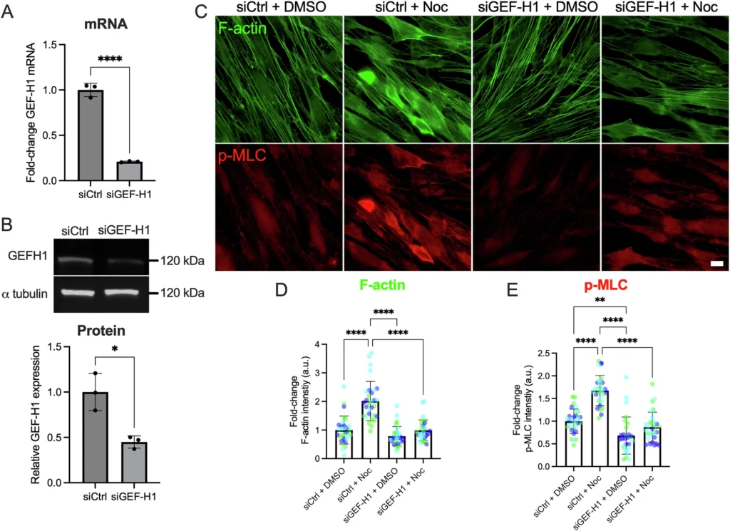
Researchers reduced GEF-H1 expression using small interfering RNA (siRNA). They then examined changes in F-actin and phosphorylated myosin light chain.
The experiments showed that GEF-H1 contributes to the relationship between microtubule stability and actomyosin activity. The results suggest that microtubules influence Schlemm’s canal cell mechanics partly through GEF-H1-related signalling.
Cell Stiffness and Transcellular Pore Formation
Another important part of the research focused on cellular stiffness. This describes how much a cell resists deformation when mechanical forces are applied.
The researchers used atomic force microscopy to measure the stiffness of Schlemm’s canal cells. Their results showed that paclitaxel treatment reduced cell stiffness compared with the control condition, while nocodazole increased it.
The team also investigated transcellular pores. These are small openings that form through Schlemm’s canal endothelial cells and allow aqueous humor to cross the inner wall of the canal.
The experiments showed that microtubule stabilisation increased particle-induced pore formation, while microtubule destabilisation reduced it. This suggests that the organisation of the cellular cytoskeleton can influence how effectively these cells form drainage openings.
In the normal-cell experiment, the graph showed pore formation increasing from approximately 9% in the control group to around 11.5% following paclitaxel treatment. The nocodazole group showed a lower result of approximately 5%.
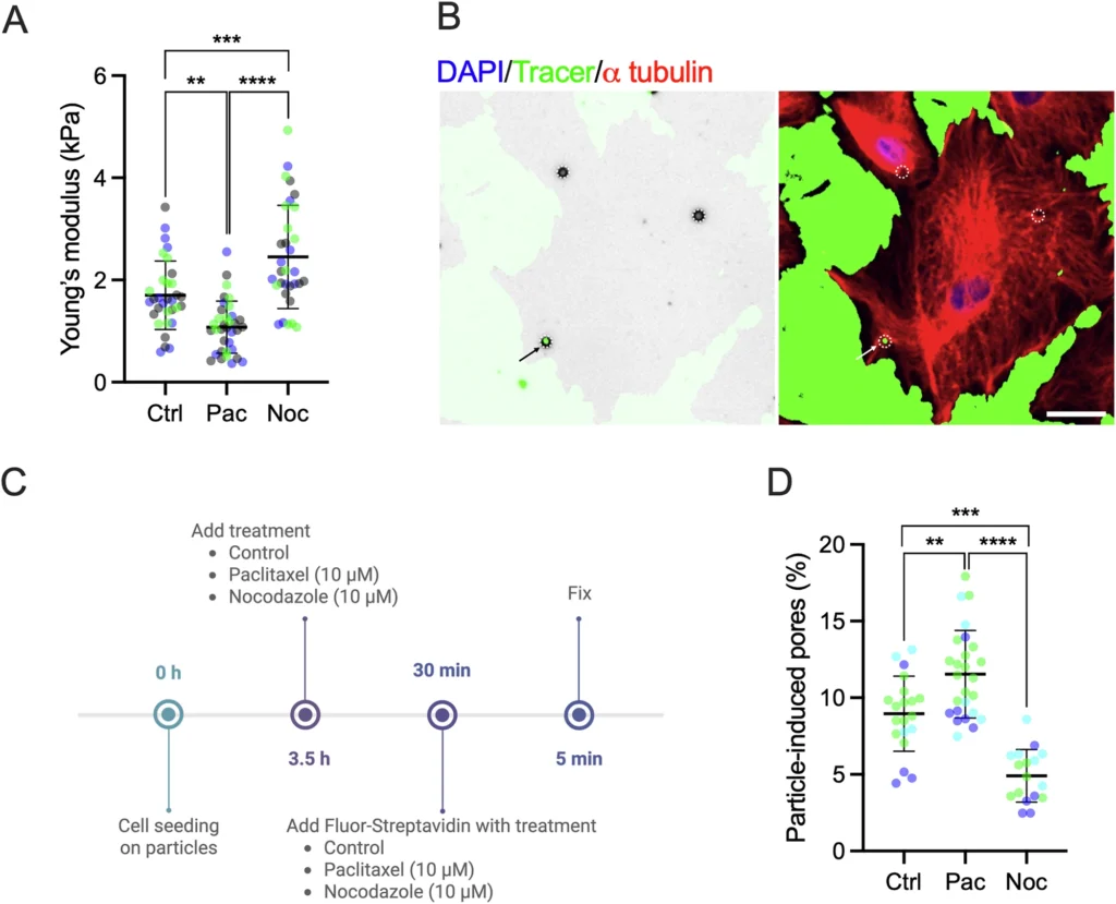
Figure 3. Microtubule stability is associated with Schlemm’s canal cell stiffness and transcellular pore formation. The experiments compare control, paclitaxel and nocodazole conditions.
A Stiffer Environment Reduces Microtubule Stability
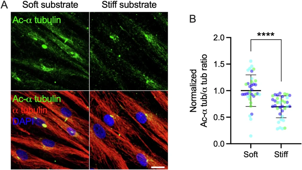
The researchers also examined how the surrounding physical environment affects microtubules. Normal Schlemm’s canal cells were grown on soft and stiff substrates to investigate whether substrate stiffness influenced their internal structure.
The results showed lower acetylated alpha-tubulin relative to total alpha-tubulin in cells grown on the stiff substrate. Acetylated alpha-tubulin is commonly used as an indicator associated with stable microtubules.
This finding suggests that the mechanical properties of the environment surrounding Schlemm’s canal cells can influence their microtubule stability.
It also provides another connection between the physical conditions inside the drainage pathway and the behaviour of the cells responsible for supporting fluid outflow.
Glaucoma-Derived Cells Show Reduced Microtubule Stability
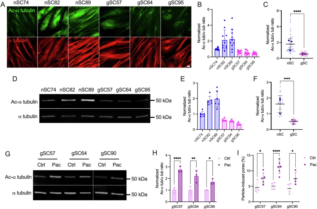
One of the most relevant findings involved the comparison between normal Schlemm’s canal cells and cells obtained from donors with glaucoma.
The researchers found that glaucoma-derived Schlemm’s canal cells displayed lower microtubule stability than normal cells. The figure compares several normal cell strains, labelled nSC, with glaucoma-derived strains, labelled gSC.
The researchers also tested whether paclitaxel could change microtubule stability in glaucoma-derived cells. Their results showed increased acetylated alpha-tubulin following treatment, indicating greater microtubule stabilisation.
Importantly, the experiments also showed increased particle-induced pore formation after paclitaxel treatment in the glaucoma-derived cell strains examined.
These findings suggest that microtubule stabilisation may influence the mechanical and drainage-related behaviour of cells obtained from glaucoma donors. However, they do not prove that reduced microtubule stability alone causes glaucoma.
Paclitaxel Increased Outflow Facility in Mouse Eyes
After examining cultured cells, the research team investigated whether microtubule stabilisation could also affect fluid drainage in whole-eye experiments.
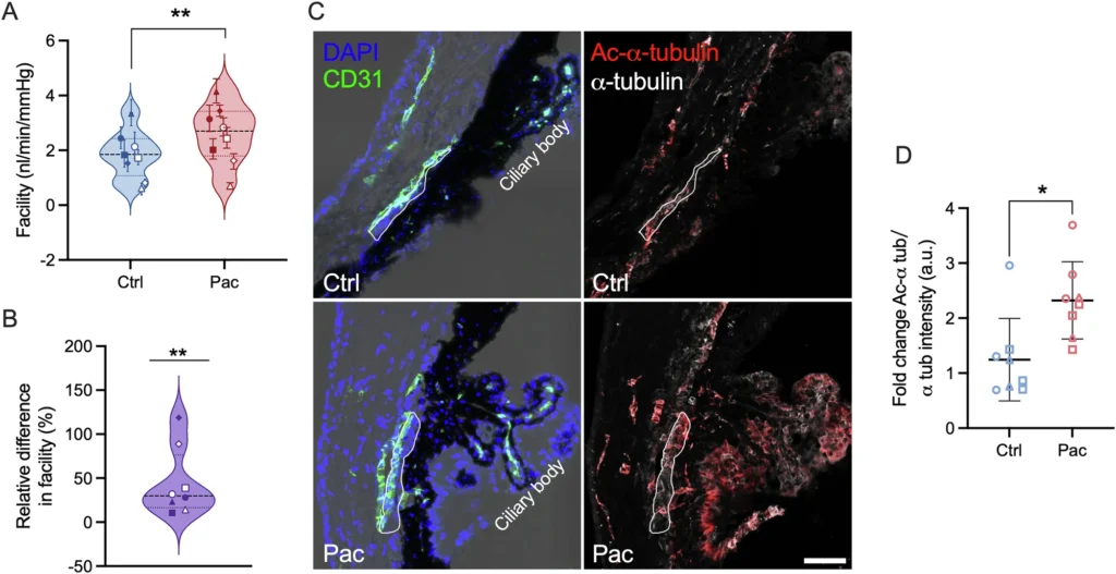
For this part, the researchers used enucleated mouse eyes and the iPerfusion system to measure aqueous humor outflow facility. Paclitaxel was used to stabilise microtubules, and the results were compared with control eyes.
The experiments showed a significant increase in outflow facility following paclitaxel perfusion. The study reported an increase of 44.3% compared with the control condition.
Outflow facility describes how easily aqueous humor moves through the conventional drainage pathway. A higher value indicates that fluid can pass through the pathway more readily.
This experiment is important because it connects the cellular findings with a measurable change in fluid drainage. However, the results came from isolated mouse eyes, not a clinical trial involving people with glaucoma.
Could This Research Lead to New Glaucoma Treatments?
The findings identify microtubule stability as a possible area for further glaucoma research. By influencing cell stiffness, contractility and pore formation, microtubules may help determine how effectively Schlemm’s canal supports aqueous humor drainage.
Current glaucoma management commonly focuses on lowering intraocular pressure through prescribed eye drops, laser procedures and surgery. These approaches aim to reduce the risk of further optic nerve damage.
Research into Schlemm’s canal cell mechanics may eventually help scientists explore additional ways to improve the conventional drainage pathway. The relationship between microtubules, GEF-H1 and cellular contractility could provide useful directions for future investigations.
However, the experiments do not establish paclitaxel as a glaucoma treatment. Its potential toxicity and effects on cell division in ocular tissues mean that further investigation is necessary before microtubule-targeting approaches could be considered for clinical use.
How Glaucoma Is Diagnosed
Glaucoma can develop without obvious early symptoms, making regular comprehensive eye examinations important. An ophthalmologist may use several tests to assess eye pressure, drainage anatomy, optic nerve health and changes in vision.
Tonometry
Tonometry measures intraocular pressure. Elevated readings may indicate an increased risk of glaucoma, although a normal measurement does not completely rule out the disease.
Gonioscopy
Gonioscopy allows an eye specialist to examine the drainage angle between the cornea and iris. It helps determine whether the angle is open or narrow and provides information about the structures involved in fluid drainage.
Optical Coherence Tomography
Optical coherence tomography, or OCT, produces detailed images of retinal structures, including the retinal nerve fibre layer. It can help identify structural changes associated with glaucoma.
Visual Field Testing
Visual field testing checks for areas of reduced peripheral vision. The results help specialists monitor functional changes that may occur as glaucoma progresses.
When Should You See an Eye Doctor?
Consider arranging a comprehensive eye examination if you have a family history of glaucoma, elevated eye pressure or previous abnormal eye test results. Do not wait for noticeable vision loss because early glaucoma may not produce obvious symptoms.
Seek urgent medical attention if you suddenly experience severe eye pain, redness, blurred vision, halos around lights, headache, nausea or vomiting. These symptoms may indicate acute angle-closure glaucoma, which requires immediate assessment.
What Researchers Need to Investigate Next
The six experimental figures provide a connected explanation of how microtubule stability relates to Schlemm’s canal cell mechanics and aqueous humor drainage. The findings cover cell contractility, GEF-H1 signalling, cellular stiffness, pore formation, differences in glaucoma-derived cells and outflow measurements in mouse eyes.
The next challenge is determining whether these mechanisms can be safely targeted in living human eyes. Further research will need to assess potential treatments, long-term effects and whether improving drainage through these pathways can help protect the optic nerve.
For people concerned about glaucoma, the practical next step remains regular eye examinations and following an ophthalmologist’s monitoring recommendations. This research offers a promising direction for understanding eye drainage, but it is not yet a clinically proven treatment.