How the Eye’s Drainage System Is Linked to Glaucoma
Glaucoma can gradually damage the optic nerve without causing noticeable symptoms in its early stages. One important factor in many cases is pressure inside the eye, which can rise when fluid does not drain properly through the eye’s natural drainage system.
A study published in Molecular Biology Reports in 2026 investigated tiny molecules called microRNAs in different regions of the human trabecular meshwork, a tissue that helps regulate fluid leaving the eye. The research examined how these molecules varied by tissue location, age and the presence of primary open-angle glaucoma.
The findings may help researchers understand the biological processes associated with aging and glaucoma. However, they do not establish a new diagnostic test or prove that the identified molecules cause the disease.
What Is the Trabecular Meshwork?
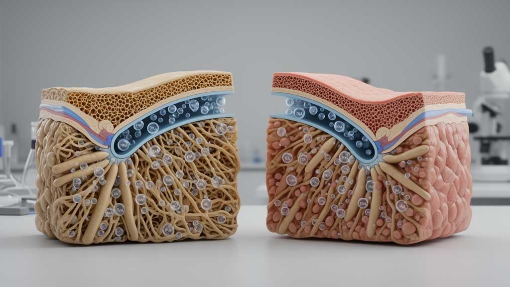
The trabecular meshwork is a network of tissue located near the junction of the cornea and iris, within the eye’s drainage angle. It helps aqueous humor, the clear fluid inside the front of the eye, flow toward the drainage channels and leave the eye.
When resistance to this outflow increases, intraocular pressure may rise. Elevated pressure is an important risk factor for glaucoma, although the disease can also develop in people whose measured eye pressure falls within the statistically normal range.
The trabecular meshwork is not uniform throughout its structure. Different regions have distinct functions and cellular characteristics, making it useful for researchers to investigate whether their molecular activity also differs.
What Did the 2026 MicroRNA Study Discover?
The research by Nair, Devarajan, Vanniarajan and colleagues, published in Molecular Biology Reports, volume 53, issue 1, Article 1472, examined microRNA expression in human donor trabecular meshwork tissue. The researchers used the nanoString nCounter platform to compare filtering and non-filtering regions.
They identified 34 microRNAs with different expression levels between the two regions, using a fold-change threshold of at least 1.2. Of these, 31 showed higher expression in the non-filtering region, while three showed lower expression compared with the filtering region.
The investigators then examined selected findings using quantitative real-time polymerase chain reaction, or qRT-PCR. This technique helps researchers measure and validate differences in RNA expression.
The study also examined donor tissue from three age groups: 0–30 years, 30–60 years and over 60 years. In addition, researchers analyzed tissue sections from donors with primary open-angle glaucoma to investigate whether particular microRNAs showed different expression patterns in glaucoma.
Seven MicroRNAs Received Further Validation
The researchers validated differences in seven microRNAs across the trabecular meshwork regions. Four showed higher expression in the non-filtering region, while three showed lower expression there.
The four with higher expression were miR-24-3p, miR-107, miR-34a-5p and miR-184. The three with lower expression were miR-376a-3p, miR-145-5p and miR-22-3p.
These seven molecules were a selected group from the original 34 differentially expressed microRNAs, rather than the complete set identified in the initial comparison.
The distinction matters because microRNAs help regulate gene activity. Understanding where their expression differs may help scientists investigate how particular cell populations maintain the tissue and how those processes change over time.
Aging Was Associated With Changes in Three MicroRNAs
One important finding involved miR-107, miR-184 and miR-34a-5p. The age-group analysis found that expression of these three molecules declined with age specifically in the non-filtering region of the trabecular meshwork.
All three had previously been validated as more highly expressed in the non-filtering region than in the filtering region. Their age-related decline therefore identifies a molecular change in a region associated with adult stem cells.
Adult stem cells help maintain and repair tissues. The study’s background describes a decline in the trabecular meshwork’s adult stem-cell population with aging and a significant reduction in primary open-angle glaucoma.
However, the researchers did not establish that declining microRNA expression directly causes the loss of these cells. Additional experiments are needed to determine whether the molecular changes influence stem-cell maintenance or simply occur alongside other age-related processes.
How Glaucoma-Associated Tissue Differed
The researchers also identified a noteworthy pattern in tissue from donors with glaucoma. The expression of miR-145-5p, miR-22-3p and miR-376a-3p was higher in glaucomatous trabecular meshwork tissue.
This finding contrasts with their lower expression in the non-filtering region compared with the filtering region in the regional analysis. The comparison suggests that tissue location and glaucoma status may be associated with different microRNA expression patterns.
It does not mean that these three molecules independently cause glaucoma. Their expression could reflect several underlying biological processes, and further investigation is required to understand their roles.
Which Biological Pathways Could Be Involved?
To explore the possible functions of the selected microRNAs, the research team used miRTarBase to investigate potential gene targets and Shiny GO 0.82 for pathway enrichment analysis.
The computational analyses associated the selected molecules with several signaling pathways, including Wnt, hypoxia-inducible factor, PI3K-AKT and MAPK signaling. These pathways participate in cellular functions such as growth, survival, responses to environmental stress and tissue maintenance.
These findings provide possible directions for future laboratory research. However, identifying a pathway through computational analysis does not prove that a particular microRNA directly regulates it in the human eye.
The study was based on donor tissue rather than an intervention in living patients. The available report also did not specify the total number of donors or provide detailed statistical estimates and uncertainty measures, which limits how broadly the findings can be interpreted.
Could MicroRNA Research Lead to Better Glaucoma Care?
In the future, research into microRNAs may help scientists identify molecular processes associated with glaucoma susceptibility, aging or changes in the eye’s drainage system. Such knowledge could eventually contribute to the development of new research tools or therapeutic approaches.
Before that becomes possible, researchers need to perform functional experiments, including microRNA transfection studies, to test how changing the levels of selected molecules affects trabecular meshwork cells.
The results would also need independent validation in additional samples. At present, the identified microRNAs should be considered research candidates rather than established clinical biomarkers or treatment targets.
Recognizing Glaucoma Symptoms and Risk Factors
Although this study explores molecular biology, people should not wait for new research developments before taking care of their eye health. Primary open-angle glaucoma often develops gradually, and early disease may cause no obvious symptoms.
As the condition progresses, peripheral vision can become impaired. Advanced glaucoma may cause substantial and irreversible vision loss, which makes appropriate screening and follow-up important.
Risk factors include increasing age, a family history of glaucoma and elevated intraocular pressure. Other factors may also affect an individual’s risk, so an eye-care professional should assess personal circumstances.
When Should You See an Eye Doctor?
Arrange a comprehensive eye examination if you have risk factors for glaucoma, have missed recommended eye checks or have been advised to monitor your eye pressure. Depending on your circumstances, an examination may include tonometry to measure intraocular pressure, gonioscopy to assess the drainage angle, optic nerve evaluation and visual field testing.
Seek urgent medical attention if you experience sudden severe eye pain, a red eye, blurred vision, halos around lights, headache or nausea. These symptoms can indicate acute angle-closure glaucoma or another urgent eye condition and should not be ignored.
What Comes Next for Glaucoma Research?
The study published in Molecular Biology Reports offers a closer look at how microRNA expression differs between trabecular meshwork regions and changes with age and glaucoma status. Its identification of 34 differentially expressed molecules, along with age-related declines in three selected microRNAs, provides a foundation for further investigation.
The next step is to establish whether these molecular patterns have direct functional effects and whether they can be reproduced in additional human samples. For readers concerned about glaucoma, the most useful action today remains scheduling appropriate eye examinations and following an eye-care professional’s recommendations rather than relying on experimental molecular findings.