What Phacolytic Glaucoma Feels Like
Imagine waking up with a red, aching eye and vision that has dimmed over a short time. Those are the classic warning signs of phacolytic glaucoma, a type of glaucoma that develops when protein leaks out of a cloudy lens.
The condition is also called lens protein glaucoma. It was first described in 1900, and eye surgeons still treat it as a serious problem today.
This article draws on a case report by Christine L. Larsen, MD, of Minnesota Eye Consultants, and Jess Boysen, MD, of the University of Minnesota Department of Ophthalmology. It appeared in the Challenging Cases column of Glaucoma Today in July/August 2015.
How Leaking Lens Protein Raises Eye Pressure
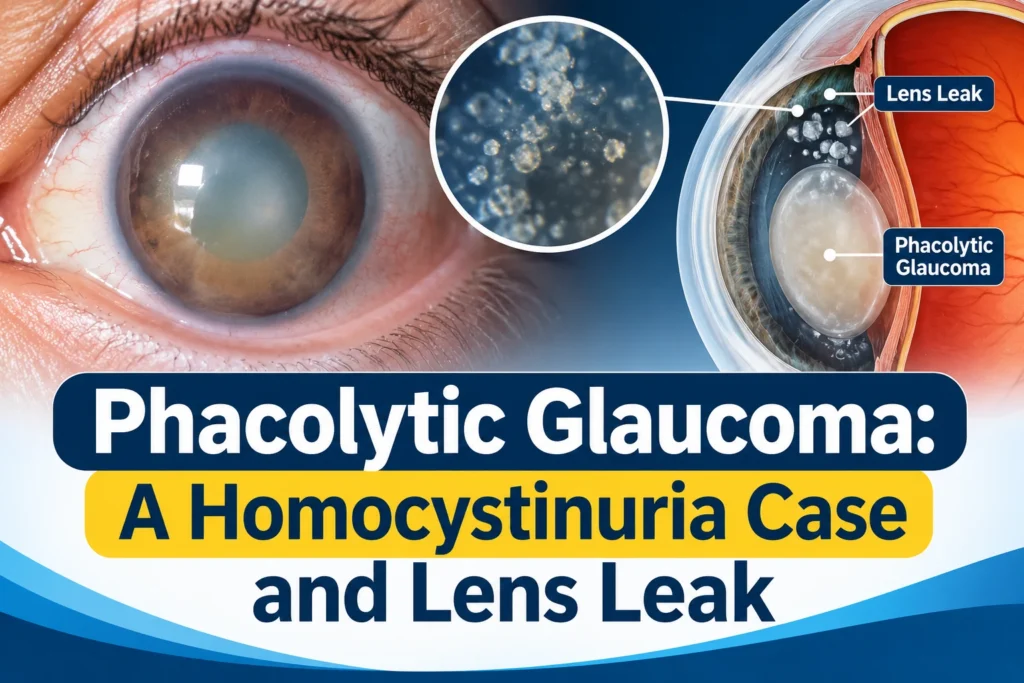
Inside the eye, the lens is wrapped in a thin capsule. In phacolytic glaucoma, soluble lens protein escapes through microscopic defects in that capsule.
The authors describe two theories for how this raises intraocular pressure (IOP). In one, macrophages that have swallowed lens material clog the trabecular meshwork, the eye’s drainage filter. In the other, the leaked protein itself blocks fluid from leaving the eye.
Doctors typically look for several signs during an exam. The angle often looks normal on gonioscopy, but heavy flare may be present. In some cases, shiny particles can be seen, which are thought to be calcium oxalate or cholesterol crystals.
A 58-Year-Old Patient With Very High Pressures
The case began when a 58-year-old man was referred for presumed aphakic glaucoma in both eyes. A routine exam at his referring doctor’s office had measured his IOP at 38 mm Hg in the right eye and 40 mm Hg in the left.
He had no eye complaints. He said he had needed a very strong glasses prescription since childhood. He was a poor historian with a very mild cognitive delay, and he reported a heart attack about 10 years earlier.
At the specialists’ office, his visual acuity was 20/80 in the right eye and 20/100 in the left, with a +10.00 D correction in both eyes. His IOP stayed in the mid-30s. The exam also showed these findings:
Front of the eye: trace conjunctival injection and 1+ anterior chamber cell in both eyes. Gonioscopy showed an angle open to the ciliary body band for 360º in both eyes.
Back of the eye: trace cells in the vitreous and optic nerve cupping of about 0.5 in both eyes. In the far inferior periphery of each eye, a dislocated crystalline lens with dense, opaque nuclear sclerotic changes was seen.
The doctors diagnosed phacolytic glaucoma. This was the less common presentation, in which the lens has dislocated into the vitreous. In those cases, the glaucoma and inflammation tend to be more subacute.
The Homocystinuria Connection
The patient had no known trauma and no sign of pseudoexfoliative material. That led the doctors to suspect a syndromic condition behind the dislocated lenses. His heart attack at a relatively young age also pointed them toward homocystinuria, and quantitative blood testing confirmed elevated homocystine.
Homocystinuria is inherited in an autosomal recessive pattern. It results from one of several enzyme deficiencies in homocysteine metabolism. Patients often develop a progressive downward lens dislocation and early myopia.
Other eye findings can include optic atrophy, iris atrophy, anterior staphylomas, and lens and corneal opacities. Body-wide features include Marfanoid habitus, pes excavatum, seizures, and thromboembolic events. The severity varies widely, depending on the enzymes involved and whether the condition responds to vitamin B6.
The authors note that newborn screening now leads to earlier diagnosis. They cite a 1989 study in the British Journal of Ophthalmology by Burke and colleagues. It reported that vitamin supplementation and/or dietary methionine restriction can slow progression and lower the risk of eye complications.
Treatment and Results
The patient was started on topical glaucoma and steroid medications. With help from a retinal specialist, he then had a vitrectomy followed by a lensectomy, along with an anterior chamber IOL implant.
After surgery, his best-corrected visual acuity improved to 20/40 in both eyes. His IOP settled in the high teens in both eyes without topical medication. His primary care provider continues to monitor his risk factors, and he is doing well.
The authors say phacolytic glaucoma is usually considered a surgical emergency. Medication is given first to reduce IOP and inflammation, followed by urgent lens removal. Patients usually regain good vision and better pressure afterward.
They also reviewed two studies on whether filtering surgery is needed. A retrospective series of 135 eyes was published in the Indian Journal of Ophthalmology in 1998 by Braganza and colleagues. In it, 89 eyes had trabeculectomy combined with cataract surgery if symptoms lasted more than 7 days or IOP stayed uncontrolled on maximum tolerated medication. The remaining eyes had cataract surgery alone.
The combined group had significantly better early IOP control. At six months, though, IOP and visual acuity were similar in both groups. In a second 1998 study by Mandal and Gothwal, in Ophthalmic Surgery and Lasers, all patients who had cataract surgery alone in a group of 45 eyes had IOP below 21 mm Hg without glaucoma medication afterward.
Based on those studies, the authors consider cataract extraction alone reasonable. They say this is especially true for recent symptoms or subacute findings.
Surgery Risks in Homocystinuria
Homocystinuria adds complexity to anesthesia. The authors note an increased risk of thromboembolic events and low blood sugar. A similar published case needed preventive measures to allow a vitrectomy under general anesthesia. Their own patient was cooperative enough for surgery under a retrobulbar blockade.
When to See a Doctor
Seek urgent eye care if you have a painful red eye with falling vision. Do not wait to see if it settles on its own. Phacolytic glaucoma is typically treated as an emergency, so quick assessment matters.
This case also shows that pressure can be very high without obvious symptoms. The patient reported no eye complaints despite IOP readings of 38 and 40 mm Hg. Regular exams that include tonometry and gonioscopy can pick up problems that you cannot feel.
Tell your eye doctor if you have had a strong glasses prescription since childhood, a known lens dislocation, or a family history of homocystinuria. Also mention any early heart or clotting problems. Your doctor can then decide whether further testing is needed.
A good next step is to book a dilated eye exam if you have not had one recently. Bring a list of your medications and your medical history. This article is general education and not a substitute for an exam.